
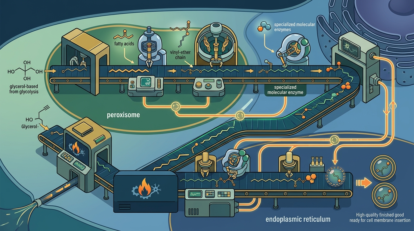
Plasmalogens do not arrive ready-made from the foods you eat. Your cells manufacture them through a precisely regulated assembly line that spans two organelles, requires at least four specialized enzymes, and depends on a feedback loop so elegant that it rivals any industrial quality-control system. This guide walks through each stage of that assembly line—from raw substrate to finished vinyl-ether lipid—and identifies the nutrients and metabolic conditions that determine whether production runs smoothly or stalls.
Starting Materials: Where the Building Blocks Come From
Every plasmalogen molecule is built on a glycerol backbone derived from glycolysis. The specific substrate is dihydroxyacetone phosphate (DHAP), an intermediate that cells produce in abundance whenever they metabolize glucose. Two additional raw materials are needed: a long-chain fatty acyl-CoA (typically C16:0 or C18:0) and a fatty alcohol generated from that acyl-CoA. The sn-2 position of the finished plasmalogen will later receive a polyunsaturated fatty acid—most often DHA (omega-3) in neurons or oleic acid (omega-9) in myelin.
Stage 1 — Inside the Peroxisome: Three Enzymes Launch the Assembly
The peroxisome is the only organelle capable of initiating plasmalogen synthesis. Three enzymes anchored to or within this organelle handle the critical opening moves.
Step 1: GNPAT Acylates DHAP
The enzyme glyceronephosphate O-acyltransferase (GNPAT) kicks off production by attaching a long-chain fatty acid to DHAP at the sn-1 position, forming 1-acyl-DHAP. This reaction takes place on the luminal side of the peroxisomal membrane. Without functional GNPAT, the entire pathway is dead on arrival.
Step 2: AGPS Creates the Ether Bond
Next, alkylglycerone phosphate synthase (AGPS) replaces the acyl group with a fatty alcohol, forging the characteristic ether linkage that distinguishes plasmalogens from ordinary phospholipids. The product is 1-O-alkyl-DHAP. AGPS works in close physical association with GNPAT, and this pairing increases catalytic efficiency.
Step 3: FAR1 Supplies the Fatty Alcohol
The fatty alcohol consumed by AGPS does not appear spontaneously. It is manufactured by fatty acyl-CoA reductase 1 (FAR1), an enzyme anchored to the cytoplasmic face of the peroxisomal membrane. FAR1 uses NADPH as a cofactor to reduce a fatty acyl-CoA into the corresponding fatty alcohol. FAR1 is widely regarded as the rate-limiting enzyme of the entire plasmalogen pathway—a point we explore further below.
Stage 2 — Completion in the Endoplasmic Reticulum
Once the alkyl-DHAP intermediate is assembled in the peroxisome, it is shuttled to the endoplasmic reticulum (ER) for four additional enzymatic steps. These ER reactions mirror the general pathway cells use to build ordinary diacyl glycerophospholipids:
- Reduction — Alkyl-DHAP is reduced to alkyl-glycerol-3-phosphate (alkyl-G3P).
- Acylation at sn-2 — A polyunsaturated fatty acid (commonly DHA or arachidonic acid) is attached at the sn-2 position, creating an alkyl-acyl phospholipid intermediate.
- Head-group addition — An ethanolamine or choline head group is added, yielding a plasmanylethanolamine or plasmanylcholine.
- Desaturation by PEDS1 — The final, defining step converts the alkyl bond into a vinyl-ether (alkenyl) bond, transforming a plasmanyl species into a true plasmalogen.
The FAR1 Bottleneck and Feedback Regulation
FAR1 does more than supply fatty alcohol; it acts as the system's master dial. Cells regulate plasmalogen levels by controlling FAR1 protein stability rather than its gene transcription. When plasmalogen concentrations in the inner leaflet of the plasma membrane rise above a set point, the cell accelerates FAR1 degradation, slowing fatty alcohol production and throttling the entire pathway. When plasmalogen levels drop, FAR1 is stabilized and production ramps up.
This feedback loop means the body can, in principle, manufacture as many plasmalogens as it needs—provided the peroxisomes are healthy and the necessary substrates and cofactors are available.
PEDS1: The Oxygen-Dependent Desaturase
The identity of the enzyme responsible for the final vinyl-ether bond was a mystery for decades. It was only recently identified as plasmanylethanolamine desaturase 1 (PEDS1), also known as TMEM189. PEDS1 requires molecular oxygen and cytochrome b5 to introduce the double bond between carbon-1 and carbon-2 of the sn-1 chain—the signature structural feature that gives plasmalogens their antioxidant reactivity and membrane-fluidity properties.
Nutrients and Cofactors That Influence Plasmalogen Production
No single supplement flips a switch on plasmalogen output, but several dietary and metabolic factors directly feed into the biosynthetic machinery.
1. Omega-3 Fatty Acids (DHA and EPA)
DHA is the predominant fatty acid at the sn-2 position of neuronal plasmalogens. Adequate dietary DHA—from fatty fish, shellfish, or algae-derived oils—ensures the ER has the right building block for brain-relevant plasmalogen species. Research has shown that EPA-enriched ethanolamine plasmalogens can increase DHA content in the brain and liver of omega-3-deficient animal models.
2. NADPH (via Niacin, Riboflavin, and Glucose Metabolism)
FAR1 is an NADPH-dependent reductase. NADPH is regenerated primarily by the pentose phosphate pathway (which requires glucose-6-phosphate dehydrogenase and vitamin B3 in the form of NAD⁺/NADP⁺) and by malic enzyme. Adequate B-vitamin status, particularly niacin (B3) and riboflavin (B2), supports the coenzyme pools that keep FAR1 functioning.
3. Choline and Ethanolamine
The head groups of plasmalogens are either ethanolamine or choline. Ethanolamine plasmalogens dominate in brain gray and white matter, while choline plasmalogens are more abundant in cardiac tissue. Dietary choline (from eggs, liver, and cruciferous vegetables) and ethanolamine (from phosphatidylethanolamine-rich foods) supply these head-group substrates.
4. B-Vitamins and Iron for Peroxisomal Health
Peroxisome biogenesis and maintenance depend on broader metabolic health. B-12, folate, and iron status influence the enzymatic landscape inside peroxisomes. These micronutrients do not directly catalyze plasmalogen steps, but deficiency in any of them can impair the organelle's overall function and, by extension, plasmalogen output.
5. Antioxidant Support
Because plasmalogens themselves are sacrificial antioxidants—their vinyl-ether bond reacts avidly with reactive oxygen species—chronic oxidative stress burns through plasmalogens faster than cells can replace them. Dietary antioxidants such as vitamin E, vitamin C, and selenium-dependent glutathione peroxidases reduce the rate of plasmalogen degradation, effectively preserving net levels even without increasing synthesis.
6. Dietary Plasmalogen Precursors
Alkylglycerols found in shark liver oil and human breast milk can bypass the peroxisomal steps and enter the ER portion of the pathway directly. While they are not raw nutrients in the traditional sense, they represent an alternative route for boosting plasmalogen levels when peroxisomal capacity is compromised.
What Happens When Biosynthesis Breaks Down
Genetic mutations in GNPAT or AGPS cause rhizomelic chondrodysplasia punctata (RCDP) types 2 and 3, severe conditions marked by skeletal malformation, cataracts, neurological impairment, and respiratory difficulties. Broader peroxisome biogenesis disorders such as Zellweger syndrome also abolish plasmalogen production because GNPAT becomes mislocalized to the cytoplasm where it is inactive.
Outside of rare genetic disease, plasmalogen levels decline naturally with aging. Reduced peroxisomal function, chronic inflammation, and cumulative oxidative stress all contribute. Lower plasmalogen levels have been documented in Alzheimer's disease, Parkinson's disease, and cardiovascular conditions—making the question of how to sustain biosynthesis a matter of practical, not just academic, importance.
Key Takeaways
- Plasmalogen biosynthesis is a two-organelle process: it begins in the peroxisome and finishes in the endoplasmic reticulum.
- Three peroxisomal enzymes—GNPAT, AGPS, and FAR1—handle the defining early steps, with FAR1 serving as the rate-limiting checkpoint.
- PEDS1 catalyzes the final oxygen-dependent desaturation that creates the vinyl-ether bond unique to plasmalogens.
- Cells self-regulate plasmalogen output through a feedback mechanism that controls FAR1 protein stability based on membrane plasmalogen concentration.
- Key nutritional inputs include DHA/EPA, NADPH-supporting B-vitamins, choline, ethanolamine, iron, and antioxidants that slow plasmalogen degradation.
- Genetic defects in any peroxisomal biosynthetic enzyme can cause devastating developmental disorders.
Frequently Asked Questions
Can you get plasmalogens directly from food?
Plasmalogens are present in shellfish, eggs, organ meats, and certain marine oils. However, the acidic environment of the stomach degrades most dietary plasmalogens before absorption. The body relies primarily on de novo synthesis in the peroxisomes rather than dietary intake to maintain tissue plasmalogen levels.
Why is FAR1 considered the rate-limiting enzyme?
FAR1 controls the supply of fatty alcohols—the critical substrate that AGPS needs to forge the ether bond. Cells regulate plasmalogen levels by adjusting FAR1 protein degradation in response to plasmalogen concentrations in the inner leaflet of the plasma membrane, making it the pathway's primary control point.
Does taking fish oil increase plasmalogen levels?
Fish oil supplies DHA, which is incorporated at the sn-2 position of neuronal plasmalogens. While DHA is a necessary building block, it does not bypass the peroxisomal steps. Adequate DHA ensures the ER has the correct fatty acid substrate, but overall plasmalogen output also depends on healthy peroxisomal function and sufficient FAR1 activity.
What role do peroxisomes play in plasmalogen production?
Peroxisomes house the first three enzymes of the plasmalogen pathway (GNPAT, AGPS, and FAR1) and are the only organelle where the characteristic ether bond can be formed. Without functional peroxisomes, plasmalogen biosynthesis cannot proceed at all, as demonstrated in peroxisome biogenesis disorders such as Zellweger syndrome.
Do plasmalogen levels decline with age?
Yes. Research consistently shows that plasmalogen concentrations decrease with advancing age. Contributing factors include reduced peroxisomal efficiency, increased oxidative stress that degrades existing plasmalogens, and chronic low-grade inflammation. This age-related decline has been linked to neurodegenerative and cardiovascular conditions.