

Your cell membranes are not static structures. They remodel themselves continuously throughout life, and one of the most consequential changes involves the slow erosion of plasmalogens—specialized ether phospholipids that protect neurons, power mitochondria, and keep inflammatory signaling in check. Understanding exactly when and how plasmalogen levels shift can help you appreciate why cellular resilience fades with time and what emerging science says about preserving it.
What Plasmalogens Actually Do in Your Membranes
Plasmalogens are a subclass of glycerophospholipids distinguished by a vinyl-ether bond at the sn-1 position of the glycerol backbone. That bond is not just a chemical curiosity—it gives the lipid a unique three-dimensional shape that influences how tightly membranes pack, how readily vesicles fuse, and how efficiently ions cross the bilayer.
Approximately 20 percent of all phospholipids in human tissue are plasmalogens, with particularly high concentrations in the brain, heart, and immune cells. In certain brain regions, plasmalogens can constitute up to nearly 90 percent of the glycerophosphoethanolamine (GPEtn) fraction. They serve roles in membrane fusion, ion transport, vesicle formation, and oxidation-reduction reactions at the membrane surface.
Their vinyl-ether bond also functions as a sacrificial antioxidant: reactive oxygen species (ROS) preferentially attack that bond, sparing neighboring polyunsaturated fatty acids from peroxidation. This protective scavenging, however, means that plasmalogens are consumed in the process—and if biosynthesis cannot keep pace, levels fall.
The Decade-by-Decade Timeline of Plasmalogen Change
Not all lipids follow the same trajectory over a human lifespan. Plasmalogen dynamics include a notable rise-then-fall pattern that diverges from the relatively stable profiles of other phospholipid classes.
Birth Through the 30s: The Building Phase
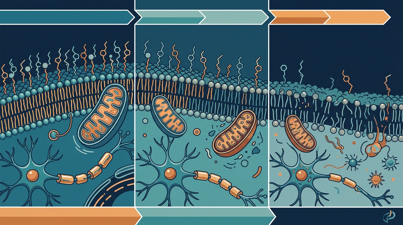
During early development and into young adulthood, the body actively accumulates plasmalogens. Myelination of the central nervous system—one of the most plasmalogen-intensive processes in biology—accelerates through childhood and continues into the mid-20s. Brain plasmalogen levels continue to climb and generally peak around the age of 30 to 40.
The 40s and 50s: The Inflection Point
After the fourth decade, a measurable decline begins. The peroxisomal biosynthetic pathway—the only route through which the body can create the ether bond characteristic of plasmalogens—starts to slow. Meanwhile, cumulative oxidative stress begins consuming plasmalogens faster than they can be replaced. Research indicates a notable decrease in plasmalogen levels particularly after the age of 50, and this decline is more pronounced in the presence of systemic inflammation.
The 60s and Beyond: Accelerating Loss
The decline steepens markedly after age 60. Classic studies by Rouser and Yamamoto, along with more recent lipidomics data, indicate that brain plasmalogen levels can fall by approximately 40 percent between the ages of 40 and 70. This is not a subtle shift—it represents a fundamental change in the biophysical properties of neuronal membranes, affecting everything from synaptic vesicle release to myelin integrity.
Four Biological Drivers Behind the Decline
The age-related drop in plasmalogens is not caused by a single mechanism. Instead, at least four converging processes contribute.
1. Peroxisomal Slowdown
Plasmalogen biosynthesis begins exclusively in peroxisomes via a nonredundant enzymatic pathway involving GNPAT and AGPS. As peroxisome number and function decline with age, the rate of new plasmalogen synthesis falls in parallel. Postmortem brain tissue from older adults and Alzheimer's patients shows decreased peroxisome density in neuronal cell bodies. Because there is no alternative biosynthetic route, any loss of peroxisomal capacity directly constrains plasmalogen supply.
2. Upregulated Degradation Enzymes
Aging is associated with increased activity of the plasmalogen-specific phospholipase A2 (PlsPE-PLA2), the enzyme responsible for cleaving plasmalogens. Higher enzyme activity means faster turnover, which compounds the supply-side deficit created by sluggish peroxisomes.
3. Chronic Oxidative Stress
The vinyl-ether bond that makes plasmalogens effective antioxidants also makes them vulnerable. Under chronic oxidative stress—common in aging tissues—reactive oxygen species degrade plasmalogens at a rate that outpaces replacement. This creates a vicious cycle: fewer plasmalogens means less antioxidant protection, which allows more ROS to accumulate, which in turn degrades plasmalogens even faster.
4. Mitochondrial Dysfunction
Recent research in Drosophila oenocytes (hepatocyte-like cells) has shown that aging drives a marked decline in mitochondrial plasmalogen levels, which correlates with impaired mitochondrial fission in response to oxidative stress. Aged cells fail to fragment enlarged mitochondria, resulting in persistently dysfunctional organelles. Genetic disruption of plasmalogen biosynthesis recapitulated this aging phenotype in young cells, suggesting that plasmalogen loss is not merely a symptom of mitochondrial decline but a causal contributor to it.
Downstream Consequences of Falling Plasmalogen Levels
Cognitive Decline and Neurodegeneration
Plasmalogen levels in the brain are abnormally low in individuals with age-related cognitive decline, dementia, and neurodegenerative diseases. Studies have found a dramatic decrease of up to 40 mol% in the plasmalogen content of white matter at early Alzheimer's disease stages. Critically, Alzheimer's patients whose serum plasmalogen levels were at or below 75 percent of age-matched controls showed significant cognitive decline over one year, while those with normal serum levels showed no measurable decline.
Synaptic Loss and Neuroinflammation
In aged rodent brains, the reduction of plasmalogen levels has been linked to both microglial dysfunction and synaptic loss. Microglia in aged brains are less active in phagocytosis, potentially due to plasmalogen depletion. Aging brains often exhibit increased neuroinflammation and synaptic loss, processes that appear to be at least partially attributable to plasmalogen deficiency. Two-month plasmalogen supplementation in aged mice alleviated hippocampal synaptic loss and promoted synaptogenesis and synaptic vesicle formation.
Membrane Remodeling and Signal Disruption
When plasmalogen levels fall, cells are forced to substitute other phospholipids—most commonly phosphatidylethanolamines—into membrane positions normally occupied by plasmalogens. This substitution changes membrane geometry, curvature, and fluidity, leading to cellular signaling abnormalities, neurotransmission deficits, and lowered antioxidant defenses.
Impaired Myelin Integrity
White matter is especially rich in plasmalogens, and different plasmalogen species are enriched in white versus grey matter. White matter plasmalogens tend to contain more saturated side chains (18:1, 20:1, 22:4), while grey matter species are enriched in polyunsaturated fatty acids such as DHA. The preferential loss of white matter plasmalogens in aging and early dementia may undermine myelin sheath stability long before clinical symptoms emerge.
Measuring Your Plasmalogen Status
One of the practical advances in plasmalogen science is the availability of blood-based measurements. While brain tissue analysis remains the gold standard for research, serum and erythrocyte plasmalogen levels have been shown to correlate with brain levels and disease severity.
Lipidomics panels that quantify ethanolamine plasmalogen (PlsEtn) species in serum can serve as accessible biomarkers. Research involving more than 400 clinically demented and 350 nondemented subjects demonstrated that circulating PlsEtn levels were significantly decreased in serum from clinically and pathologically diagnosed Alzheimer's subjects at all stages of dementia. Erythrocyte plasmalogen levels have also been shown to correlate with disease severity, supporting the idea that plasmalogen depletion reflects a systemic—not merely cerebral—process.
Emerging Strategies for Preservation and Restoration
While no intervention can fully reverse age-related peroxisomal decline, several strategies are under active investigation.
Plasmalogen Replacement Therapy
Plasmalogen replacement therapy (PRT) has been shown in preclinical models to restore plasmalogen levels and ameliorate pathological phenotypes. In aged mice, oral plasmalogen supplementation enhanced synaptic plasticity, promoted neurogenesis in the hippocampus, and inhibited age-related microglial activation.
Oral Plasmalogen Supplementation in Humans
A multicenter, randomized, double-blind, placebo-controlled trial published in eBioMedicine found that oral administration of scallop-derived purified plasmalogens may improve cognitive functions in patients with mild Alzheimer's disease. However, study durations have been relatively short, and larger-scale trials are needed to confirm long-term efficacy.
Supporting Peroxisomal Health
Because the peroxisome is the bottleneck in plasmalogen synthesis, strategies that support peroxisomal biogenesis and function—including adequate DHA intake, regular physical activity, and reducing chronic inflammatory load—may help maintain endogenous production. Research on n-3 fatty acid intake and plasmalogen metabolism-related enzymes in the aging brain continues to explore this connection.
Always consult a qualified healthcare provider before starting any supplementation regimen.
Key Takeaways
- Brain plasmalogen levels rise through early adulthood, peak around age 30–40, and can decline by roughly 40 percent by age 70.
- Peroxisomal slowdown, increased degradation enzyme activity, chronic oxidative stress, and mitochondrial dysfunction all contribute to the decline.
- Low plasmalogen levels are strongly associated with cognitive decline, neurodegeneration, synaptic loss, and neuroinflammation.
- Blood-based lipidomics can measure plasmalogen status and may serve as an early biomarker for dementia risk.
- Plasmalogen replacement therapy shows promise in animal models and early human trials, but more research is needed.
- Supporting peroxisomal health through lifestyle and nutrition may help preserve endogenous plasmalogen production.
Frequently Asked Questions
At what age do plasmalogen levels start to decline?
Research indicates that brain plasmalogen levels peak around age 30–40 and begin declining thereafter, with a particularly notable decrease after age 50. The decline accelerates after 60, and by age 70, brain levels may have fallen by approximately 40 percent compared to their peak.
Can plasmalogen loss be measured with a blood test?
Yes. Serum ethanolamine plasmalogen (PlsEtn) levels have been validated as a peripheral biomarker in research involving hundreds of subjects. Erythrocyte plasmalogen levels also correlate with brain levels and dementia severity, making blood-based lipidomics a practical screening approach.
Do all tissues lose plasmalogens at the same rate?
No. The brain and heart, which have the highest baseline concentrations, are especially vulnerable. Within the brain, white matter tends to show earlier and more severe depletion than grey matter. The rate of loss also depends on local oxidative stress levels, peroxisome density, and inflammatory environment.
Is plasmalogen decline a cause or consequence of Alzheimer's disease?
Evidence supports a bidirectional relationship. Age-related peroxisomal decline reduces plasmalogen supply independently of Alzheimer's pathology, but Alzheimer's-associated oxidative stress and inflammation further accelerate degradation. Some researchers have proposed peripheral ethanolamine plasmalogen deficiency as a logical causative factor in Alzheimer's disease and dementia, not merely a downstream effect.
Can supplements restore plasmalogen levels?
Preclinical studies and early clinical trials suggest that oral plasmalogen supplementation can raise circulating levels and may improve cognitive outcomes. However, direct plasmalogen supplements remain relatively expensive, and long-term human data is still limited. Discuss any supplementation strategy with your healthcare provider.
What role do peroxisomes play in plasmalogen production?
Peroxisomes are the exclusive site where the vinyl-ether bond of plasmalogens is formed. Key enzymes including GNPAT, AGPS, and DHAPAT reside in peroxisomes. As peroxisomal function declines with age, plasmalogen biosynthetic capacity decreases proportionally, because there is no alternative metabolic pathway to compensate.