

Your cells build every plasmalogen molecule from scratch. The process spans two distinct organelles, relies on a handful of specialized enzymes, and is governed by an elegant feedback loop that keeps supply in balance with demand. Here is a science-grounded look at each stage of the pathway—and the dietary nutrients that can influence whether it functions at its best.
Why De Novo Synthesis Matters More Than Diet Alone
Although certain animal foods—especially seafood and organ meats—contain preformed plasmalogens, the body cannot rely on dietary intake alone. De novo (from-scratch) biosynthesis is the primary mechanism for maintaining adequate levels in tissues. Research on peroxisome biogenesis disorders has demonstrated this convincingly: patients whose peroxisomes cannot function properly show plasmalogen levels in brain, heart, and kidney that drop below 10% of normal values, despite normal dietary intake. This confirms that the body's own synthetic machinery is the dominant source of tissue plasmalogens.
Phase 1 — The Peroxisome Phase
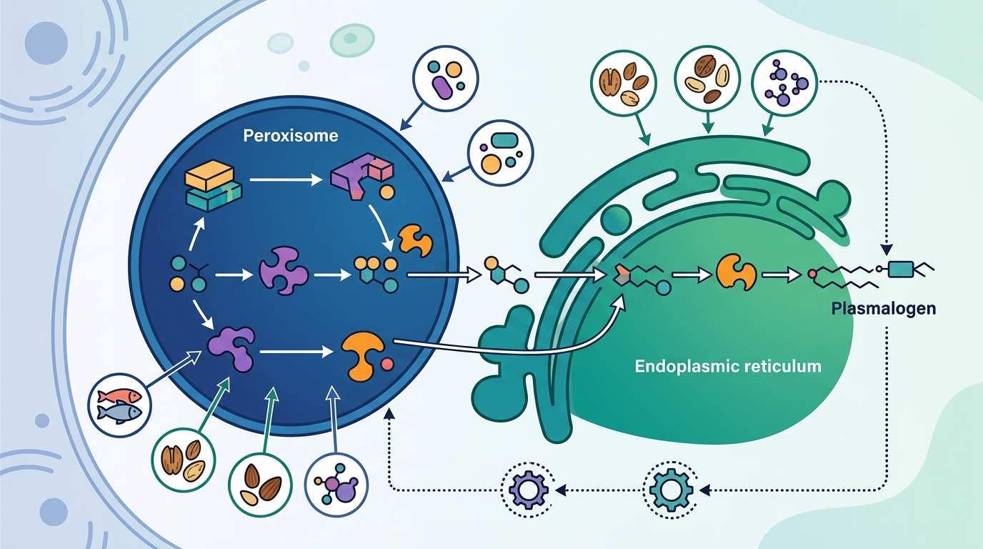
Plasmalogen biosynthesis begins inside peroxisomes, small oxidative organelles present in virtually every cell type. Two matrix enzymes carry out the critical opening reactions:
Step 1: GNPAT Acylates DHAP
The enzyme glyceronephosphate O-acyltransferase (GNPAT) kicks off the pathway. It attaches a fatty acyl chain to dihydroxyacetone phosphate (DHAP) at the sn-1 position, creating acyl-DHAP. This is the committed first step; without functional GNPAT, no downstream intermediates can form.
Step 2: AGPS Introduces the Ether Bond
Next, alkylglycerone phosphate synthase (AGPS) swaps the acyl chain for a long-chain fatty alcohol, creating alkyl-DHAP. This reaction introduces the characteristic ether linkage that distinguishes plasmalogens from ordinary phospholipids. GNPAT and AGPS form a heterotrimeric complex inside the peroxisome, which is believed to facilitate substrate channeling between the two reactions.
The Role of FAR1 on the Peroxisomal Membrane
The fatty alcohol that AGPS needs does not appear spontaneously. It is produced by fatty acyl-CoA reductase 1 (FAR1), a C-tail-anchored protein on the cytoplasmic face of the peroxisomal membrane. FAR1 uses NADPH to reduce a fatty acyl-CoA into the corresponding fatty alcohol, which then enters the peroxisomal matrix for AGPS to use.
Step 3: AADHAP-R Reduces the Intermediate
The third enzyme, acyl/alkyl-DHAP reductase (AADHAP-R), is found at both peroxisomal and ER membranes. It reduces the ketone at the sn-2 position of alkyl-DHAP, generating 1-alkyl-2-lyso-sn-glycero-3-phosphate. This product is then shuttled to the endoplasmic reticulum for the remaining steps.
Phase 2 — The Endoplasmic Reticulum Phase
Once the alkyl-glycerophosphate intermediate reaches the ER, several additional enzymes complete the construction of the plasmalogen precursor:
- Acyltransferase (AAG3P-AT) attaches a second fatty acyl chain—often an omega-3 or omega-6 polyunsaturated fatty acid such as DHA or arachidonic acid—at the sn-2 position.
- Phosphohydrolase (PH) removes the phosphate group from the sn-3 position.
- Ethanolamine phosphotransferase (E-PT) adds the ethanolamine headgroup via a CDP-ethanolamine intermediate, producing plasmanylethanolamine—the immediate precursor of the finished plasmalogen.
Altogether, this sequence from peroxisome to ER involves seven enzymatic reactions, a figure consistently reported across the peer-reviewed literature.
The Final Step: TMEM189 and the Vinyl-Ether Bond
The reaction that actually makes a plasmalogen a plasmalogen—introduction of the vinyl-ether double bond at sn-1—was a mystery until 2020. Researchers identified the gene TMEM189 as encoding plasmanylethanolamine desaturase (PEDS), the integral ER membrane protein responsible for inserting this bond. Inactivation of TMEM189 in human HAP1 cells caused a total loss of PEDS activity and a 17-fold reduction in plasmalogen levels, while plasmanylethanolamine substrates accumulated upstream. This study, published in Proceedings of the National Academy of Sciences (Werner et al., 2020), settled a decades-long search for the enzyme that creates the hallmark structural feature of plasmalogens.
The FAR1 Feedback Loop — Built-In Quality Control
Cells do not produce plasmalogens blindly. A sophisticated feedback mechanism ensures that production matches demand. At the center of this loop is FAR1, the enzyme that supplies fatty alcohols for the peroxisomal steps.
Here is how the loop works:
- Finished plasmalogens are transported to the inner leaflet of the plasma membrane.
- A sensing mechanism detects the concentration of plasmalogens in that inner leaflet.
- When levels are sufficient, FAR1 protein on the peroxisomal membrane is targeted for accelerated degradation, slowing the supply of fatty alcohols and throttling the entire pathway.
- When levels drop, FAR1 degradation slows, FAR1 protein accumulates, and biosynthesis speeds up.
This means FAR1 acts as both the rate-limiting enzyme and the primary regulatory node for the entire pathway. The discovery of this mechanism has important implications: any factor that impairs peroxisomal function, disrupts membrane sensing, or accelerates plasmalogen degradation (such as chronic oxidative stress) can throw the feedback loop out of balance.
Post-Synthesis Transport
After synthesis is complete, plasmalogens must reach their functional destinations—primarily cell membranes throughout the body. They are transported to post-Golgi compartments, including endosomes and the plasma membrane, through an ATP-dependent but non-vesicular pathway. Once at the plasma membrane, a P4-type ATPase called ATP8B2 helps maintain their preferential localization in the inner leaflet, the position from which the feedback loop senses their concentration.
Nutrients That Support Plasmalogen Production
The biosynthetic pathway has specific metabolic requirements. Although no single nutrient can overcome a genetic defect in peroxisome function, the following dietary factors supply substrates or cofactors for enzymes along the pathway:
| Nutrient Category | Role in Plasmalogen Biosynthesis | Dietary Sources |
|---|---|---|
| Omega-3 fatty acids (DHA, EPA) | Incorporated at sn-2 position of the glycerol backbone; critical for brain-type ethanolamine plasmalogens | Salmon, mackerel, sardines, algae oil, flaxseeds |
| Omega-6 fatty acids (arachidonic acid) | Alternative sn-2 acyl chain in many tissue-specific plasmalogens | Nuts, seeds, vegetable oils, eggs |
| Choline | Required for choline-type plasmalogens (PlsCho); a headgroup precursor | Eggs, liver, lean meats, cruciferous vegetables |
| Ethanolamine | Headgroup precursor for the dominant ethanolamine-type plasmalogens (PlsEtn) | Meats, fish, eggs, dairy |
| NADPH (via niacin / B3) | Cofactor for FAR1 (fatty alcohol production) and AADHAP-R (reduction step) | Whole grains, poultry, fish, legumes |
| B vitamins (B2, B6, B12, folate) | Support methylation and one-carbon metabolism that intersects with phospholipid remodeling | Meats, eggs, leafy greens, legumes |
| Vitamin E (alpha-tocopherol) | Fat-soluble antioxidant that helps protect the oxidation-sensitive vinyl-ether bond from premature cleavage | Almonds, sunflower seeds, spinach, avocado |
| Vitamin C | General antioxidant defense; note that in vitro evidence suggests high concentrations can cleave the vinyl-ether bond, so balance matters | Citrus fruits, bell peppers, strawberries |
| Selenium | Cofactor for glutathione peroxidases, which reduce lipid peroxides that can degrade plasmalogens | Brazil nuts, seafood, organ meats |
Importantly, the pathway also depends on healthy peroxisome biogenesis, adequate mitochondrial energy production (for ATP-dependent transport), and manageable levels of oxidative stress. A nutrient-dense, anti-inflammatory dietary pattern supports all three of these conditions simultaneously.
What Goes Wrong: Conditions Linked to Impaired Biosynthesis
Understanding the pathway makes it easier to see how disruptions lead to disease:
- Rhizomelic chondrodysplasia punctata (RCDP) — Caused by mutations in genes encoding GNPAT, AGPS, or the peroxisomal import factor PEX7. Results in a near-total absence of plasmalogens and severe developmental abnormalities.
- Zellweger spectrum disorders — Peroxisome biogenesis defects that mislocalize GNPAT to the cytoplasm, where it cannot function, leading to profoundly low plasmalogen levels across all organs.
- Age-related decline — Plasmalogen levels fall with aging. Whether this reflects reduced peroxisomal function, increased oxidative degradation, or both remains an area of active investigation.
- Alzheimer's disease — Reduced ethanolamine plasmalogen levels have been documented in Alzheimer's patients, and TMEM189 (the gene for the final desaturase step) is among the genes of interest in understanding this lipid deficit.
Key Takeaways
- Plasmalogen biosynthesis is a seven-step process that starts in peroxisomes (GNPAT → AGPS → AADHAP-R) and finishes in the endoplasmic reticulum (acyltransferase → phosphohydrolase → ethanolamine phosphotransferase → TMEM189/PEDS).
- FAR1 on the peroxisomal membrane is the rate-limiting enzyme and is regulated by a negative feedback loop that senses plasmalogen concentration in the inner leaflet of the plasma membrane.
- TMEM189, identified in 2020, encodes the desaturase that introduces the vinyl-ether bond—the structural signature of all plasmalogens.
- Omega-3 and omega-6 fatty acids supply the sn-2 acyl chains; ethanolamine and choline supply the headgroups; B vitamins and NADPH support enzymatic cofactor needs.
- Antioxidant nutrients (vitamin E, selenium) protect the oxidation-sensitive vinyl-ether bond from premature degradation.
- Healthy peroxisome function is non-negotiable—genetic or metabolic disruptions to peroxisome biogenesis can reduce tissue plasmalogens to below 10% of normal.
Frequently Asked Questions
Where in the cell are plasmalogens made?
Plasmalogen biosynthesis begins in peroxisomes, where the first two to three enzymatic steps occur, and is completed in the endoplasmic reticulum (ER) through four additional steps. The finished molecules are then transported to the plasma membrane and other cellular compartments.
What is the rate-limiting enzyme in plasmalogen biosynthesis?
Fatty acyl-CoA reductase 1 (FAR1) is considered the rate-limiting enzyme. It sits on the peroxisomal membrane and produces the fatty alcohols needed for the ether bond. Its protein levels are regulated through a feedback loop that senses how many plasmalogens are present in the plasma membrane.
What enzyme creates the vinyl-ether bond in plasmalogens?
Plasmanylethanolamine desaturase (PEDS), encoded by the TMEM189 gene, catalyzes the introduction of the vinyl-ether double bond at the sn-1 position. This is the final step of plasmalogen biosynthesis and takes place in the ER membrane.
Can I boost plasmalogen levels through diet?
Diet can support—but not single-handedly determine—plasmalogen levels. Omega-3 fatty acids, choline, ethanolamine, B vitamins, and antioxidant nutrients like vitamin E all contribute substrates or cofactors to the biosynthetic pathway. However, because de novo synthesis is the primary source of tissue plasmalogens, peroxisomal health and overall metabolic function matter just as much as what you eat.
Why do plasmalogen levels decline with age?
The exact mechanism is still under investigation. Contributing factors likely include reduced peroxisomal function, increased oxidative stress that degrades the vulnerable vinyl-ether bond, and age-related changes in the FAR1 feedback loop. Chronic inflammation may also accelerate plasmalogen turnover beyond the body's capacity to replenish them.