

Why do certain lipids vanish from the brain years before a diagnosis of Alzheimer's or Parkinson's disease? This guide traces the journey of plasmalogens — from their synthesis inside peroxisomes to the clinical observations that now place them at the center of neurodegeneration research.
What Are Plasmalogens and Why Does the Brain Need Them?
Plasmalogens are a subclass of ether phospholipids distinguished by a vinyl-ether bond at the sn-1 position and polyunsaturated fatty acids — commonly docosahexaenoic acid (DHA) — at sn-2. They represent roughly one in five phospholipids in human tissue and are particularly concentrated in the brain, heart, and immune cells.
Within the central nervous system, plasmalogens perform several overlapping roles: they modulate membrane fluidity, facilitate vesicle fusion during neurotransmitter release, serve as reservoirs for signaling lipids such as arachidonic acid and DHA, and act as endogenous antioxidants through their reactive vinyl-ether linkage.
The brain contains the highest concentration of plasmalogens of any organ, which is why even modest declines in plasmalogen levels can create downstream effects on cognition, signaling, and cellular resilience.
Peroxisomal Biosynthesis: Where Plasmalogens Begin
Plasmalogen synthesis is initiated exclusively inside peroxisomes — small, membrane-bound organelles that also handle fatty acid beta-oxidation and reactive oxygen species management. The first two enzymatic steps, catalyzed by dihydroxyacetone phosphate acyltransferase (DHAP-AT) and alkyl-DHAP synthase, occur on the inner peroxisomal membrane. The intermediate is then shuttled to the endoplasmic reticulum for completion.
This strict peroxisomal dependency means that anything impairing peroxisome biogenesis — genetic mutations, aging-related organelle decline, or chronic oxidative burden — directly curtails plasmalogen production. In peroxisomal biogenesis disorders such as Zellweger spectrum, patients present with severe plasmalogen deficiency and profound neurological impairment from birth, underscoring how critical this biosynthetic pathway is for the nervous system.
Quantifying the Loss: Lipidomics Data in AD and PD
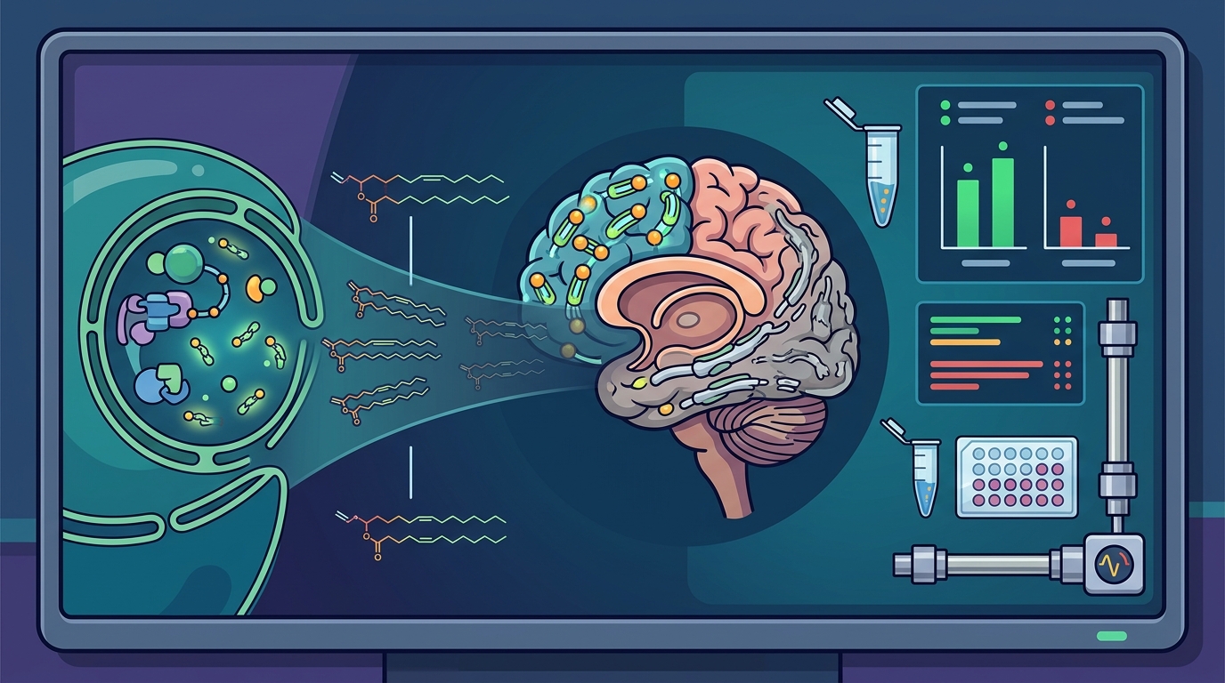
Modern lipidomics platforms have enabled precise measurement of plasmalogen subspecies across biological compartments. The findings in Alzheimer's disease are striking: shotgun lipidomics studies have demonstrated a dramatic decrease in ethanolamine plasmalogen content in white matter at the very mild stage of AD, with gray matter depletion correlating with disease severity — from roughly 10 mol% loss in very mild cases to 30 mol% reduction in severe cases.
Reductions in ethanolamine plasmalogens (PlsEtns) have been reported in plasma, serum, cerebrospinal fluid, and brain tissue of AD patients, leading researchers to propose PlsEtns as a candidate biomarker for the disease.
In Parkinson's disease, reduced PlsEtn levels have been documented across multiple independent studies. Reduced levels of PlsEtns have been found to be associated with aging and several neurodegenerative diseases, including Parkinson's disease, Niemann-Pick type C disease, multiple sclerosis, and Zellweger syndrome, establishing plasmalogen loss as a shared feature of neurodegeneration rather than a disease-specific anomaly.
Myelin, White Matter, and the Structural Consequences of Depletion
Myelin sheaths — the insulating layers around axons that allow rapid signal conduction — are among the most plasmalogen-rich structures in the body. Ethanolamine plasmalogens can constitute over 80% of certain myelin phospholipid fractions. When plasmalogen levels fall, myelin integrity deteriorates.
In animal models of plasmalogen deficiency, selective knockout of oligodendrocyte peroxisomes causes white matter plasmalogen defects, morphological changes, and symptomology similar to multiple sclerosis. This observation reinforces the idea that impaired plasmalogen availability compromises the very architecture of neural signal transmission.
In Alzheimer's disease, the early and severe depletion of white-matter plasmalogens may explain some of the connectivity failures that precede frank neuronal death — a finding that reframes AD in part as a disorder of membrane lipid maintenance rather than purely a proteinopathy.
Oxidative Stress: The Vinyl-Ether Shield Theory
The vinyl-ether bond at sn-1 is more chemically reactive than the ester bonds found in conventional phospholipids. This reactivity is a double-edged sword: it makes plasmalogens susceptible to oxidative cleavage, but this same property allows them to function as sacrificial antioxidants that scavenge reactive oxygen species (ROS) before those radicals can damage proteins or DNA.
In a clinical trial involving cognitively impaired persons, plasmalogen precursor supplementation improved oxidative stress biomarkers including malondialdehyde, catalase, and superoxide dismutase, with improvements correlating with higher DHA-plasmalogen levels. This suggests that replenishing plasmalogens may restore a layer of endogenous antioxidant defense that is lost during neurodegeneration.
From a mitochondrial perspective, elevated ROS output from aging or dysfunctional mitochondria accelerates plasmalogen consumption, creating a vicious cycle: oxidative stress depletes plasmalogens, and reduced plasmalogen levels remove a key antioxidant buffer, allowing further oxidative damage.
Synaptic Health and Microglia-Mediated Neuroinflammation
A 2022 study published in Frontiers in Molecular Biosciences provided compelling evidence that plasmalogen supplementation can reverse aging-related synaptic damage. In aged mice receiving two months of intragastric plasmalogen administration, transmission electron microscopy revealed alleviation of age-associated hippocampal synaptic loss and promotion of synaptogenesis and synaptic vesicle formation. RNA sequencing and immunofluorescence analyses confirmed that plasmalogens remarkably enhanced both synaptic plasticity and neurogenesis in the aged hippocampus.
The same study demonstrated anti-neuroinflammatory effects, with plasmalogens reducing microglia-mediated inflammation. Because chronic microglial activation is implicated in both Alzheimer's and Parkinson's pathology, this finding positions plasmalogens as molecules that act at the intersection of structural repair and immune modulation within the brain.
RCDP and the Causal Evidence for Plasmalogen-Linked Neurodegeneration
One of the strongest arguments for a causal — rather than merely correlational — relationship between plasmalogen deficiency and neurodegeneration comes from rhizomelic chondrodysplasia punctata (RCDP), a genetic disorder that severely impairs peroxisomal plasmalogen synthesis.
Patients with RCDP present with myelination deficits, enlarged ventricles, subarachnoid space expansion, and cerebellar atrophy — the latter attributed to loss of Purkinje cells. This constellation of pathology mirrors aspects of the neurodegeneration seen in AD and PD.
In Pex7 knockout mouse models of RCDP, supplementation with alkyl glycerol plasmalogen precursors before pathology onset ameliorated nerve conduction deficits and reduced neurodegeneration, providing experimental evidence on the causal association of plasmalogens and neurodegeneration. A 2019 analysis concluded that plasmalogen deficiency appears to meet Bradford Hill criteria for causal association with neurodegeneration to a considerable extent.
Biomarker Potential: Early Detection Before Cognitive Decline
Perhaps the most clinically urgent implication of plasmalogen research is the possibility of using plasmalogen levels as an early biomarker. Alzheimer's pathology begins well before symptoms are manifested. When the loss of neuronal cells reaches a critical point with cognition deficit, approximately 50 to 80 percent of cells have already died — a stage at which reversing disease progression becomes extremely difficult.
Blood-based plasmalogen measurements are non-invasive and relatively inexpensive compared to PET imaging or cerebrospinal fluid taps. The consistent observation of reduced PlsEtns across plasma, serum, CSF, and brain tissue suggests that peripheral measurements may reflect central nervous system status with sufficient fidelity for screening purposes.
Research groups are actively developing standardized assays that could be deployed in routine clinical settings, though consensus on cut-off values and age-adjusted reference ranges is still pending.
Supplementation and Emerging Therapeutic Strategies
Plasmalogen Precursors
Oral administration of 1-O-alkyl-2-acylglycerol (AAG) plasmalogen precursors containing DHA at the sn-2 position has been shown to dose-dependently increase blood DHA plasmalogens and demonstrate neuroprotective effects in animal models at doses between 10 and 50 mg/kg. An investigational clinical trial in 22 cognitively impaired persons using an escalating dosing regimen of DHA-AAG from 900 to 3,600 mg/day over four months observed statistically significant improvement in cognition and mobility.
Lipid Nanoparticle Delivery
Emerging research into plasmalogen-based lipid nanoparticles (LNPs) represents a frontier in targeted delivery. Plasmalogen-based nanoassemblies — including vesicles, hexosomes, and cubosomes — have been shown to control the kinetics of CREB activation in a sustained manner and are proposed to provide more effective treatments for neurodegenerative disorders. In a transgenic Parkinson's disease mouse model, LNP-delivered plasmalogens demonstrated advantages over conventional delivery. These nanoformulations also upregulate ERK-Akt signaling pathways in neuronal cells, enhancing recruitment of the CREB transcription factor to the BDNF promoter region — a pathway critical for neuronal survival and plasticity.
Dietary and Lifestyle Considerations
Marine-derived foods — particularly shellfish, certain fish species, and sea squirts — are among the richest dietary sources of plasmalogens. Because plasmalogen levels decline with both age and chronic stress, lifestyle factors that support peroxisomal health — including regular physical activity, adequate sleep, and reduction of chronic inflammatory triggers — may help preserve endogenous synthesis capacity.
Key Takeaways
- Plasmalogens are ether phospholipids enriched in brain tissue, making up approximately one in five phospholipids in the human body and playing critical roles in membrane structure, antioxidant defense, and synaptic signaling.
- Lipidomics studies show plasmalogen depletion begins at the very mild stage of Alzheimer's disease and correlates with severity — losses range from 10 to 30 mol% in gray matter depending on disease stage.
- Reduced ethanolamine plasmalogens are documented in Alzheimer's disease, Parkinson's disease, multiple sclerosis, and genetic peroxisomal disorders, suggesting a shared mechanism of neurodegeneration.
- RCDP — a genetic disorder of plasmalogen synthesis — produces neurodegeneration that mirrors aspects of AD and PD, providing evidence for a causal rather than merely correlational relationship.
- Plasmalogen precursor supplementation has improved oxidative stress markers, cognition, and mobility in cognitively impaired persons in early clinical trials.
- Plasmalogen-based lipid nanoparticles represent an emerging therapeutic strategy that can sustain CREB signaling activation in neurodegenerative disease models.
- Blood plasmalogen levels hold promise as a non-invasive early biomarker for neurodegeneration, potentially enabling intervention before irreversible neuronal loss occurs.
Frequently Asked Questions
What are plasmalogens and why are they important for the brain?
Plasmalogens are a class of ether phospholipids characterized by a vinyl-ether bond at the sn-1 position. They are found in especially high levels in neuronal membranes, where they support membrane fluidity, neurotransmitter release, and antioxidant defense. The brain contains the highest concentration of plasmalogens of any organ.
How are plasmalogen levels measured?
Plasmalogen levels are measured using lipidomics techniques, including shotgun lipidomics and liquid chromatography–mass spectrometry (LC-MS). Measurements can be taken from blood serum, plasma, cerebrospinal fluid, or brain tissue. Blood-based assays offer a non-invasive screening option.
Do plasmalogen levels decline with age?
Yes. Plasmalogen levels naturally decrease with aging. This age-related decline is accelerated by chronic stress and oxidative burden and may contribute to increased vulnerability to neurodegenerative diseases.
Is there evidence that plasmalogen deficiency causes neurodegeneration?
Genetic disorders like RCDP, which severely impair plasmalogen synthesis, produce profound neurodegeneration from birth — including myelination deficits and cerebellar atrophy. Researchers have noted that plasmalogen deficiency meets Bradford Hill criteria for causal association with neurodegeneration. In animal models, supplementation with plasmalogen precursors before pathology onset prevented neurodegeneration.
Can plasmalogen supplements help with Alzheimer's or Parkinson's disease?
Early-stage clinical and preclinical evidence is encouraging. An investigational trial showed that DHA-containing plasmalogen precursors improved cognition, mobility, and oxidative stress markers in cognitively impaired persons. Lipid nanoparticle delivery systems are also being investigated for targeted plasmalogen delivery in Parkinson's disease models. However, large-scale randomized controlled trials are still needed.
What foods contain plasmalogens?
Marine-derived foods such as shellfish, certain fatty fish, and sea squirts (ascidians) are among the richest dietary sources of plasmalogens. Animal-derived foods including organ meats also contain meaningful amounts.