

Plasmalogens — the vinyl-ether phospholipids woven into nearly every cell membrane in your body — are finally getting the scientific attention they deserve. But how do laboratories actually detect and quantify these lipids, and which biomarkers matter most for understanding your health? This guide breaks down every major analytical platform, the specific molecular species researchers track, and how clinical testing is evolving to bring plasmalogen science from bench to bedside.
Why Measure Plasmalogens at All?
Plasmalogen levels are not static. They shift with age, oxidative burden, peroxisomal function, and disease state. Reduced circulating plasmalogens are increasingly recognized as a clinically meaningful biomarker. According to research published in the Journal of Lipid Research, a reduction of circulating plasmalogens is increasingly used clinically as a biomarker of disease, with altered plasmalogen metabolism described across chronic cardiac, hepatic, renal, and systemic conditions. Because plasmalogens are synthesized in the peroxisomes and completed at the endoplasmic reticulum, their blood concentration reflects the functional integrity of an entire biosynthetic chain.
Measuring them gives clinicians and researchers a window into membrane health, oxidative stress resilience, and peroxisomal competence — three pillars of cellular vitality that standard lipid panels completely ignore.
Analytical Platforms for Plasmalogen Quantification
The challenge with measuring plasmalogens is that their vinyl-ether bond makes them chemically distinct from — yet structurally similar to — other ether lipids. The analytical method must differentiate plasmenyl (P-) species from plasmanyl (O-) species to produce accurate results. Below are the principal technologies used in modern laboratories.
1. Liquid Chromatography–Tandem Mass Spectrometry (LC-MS/MS)
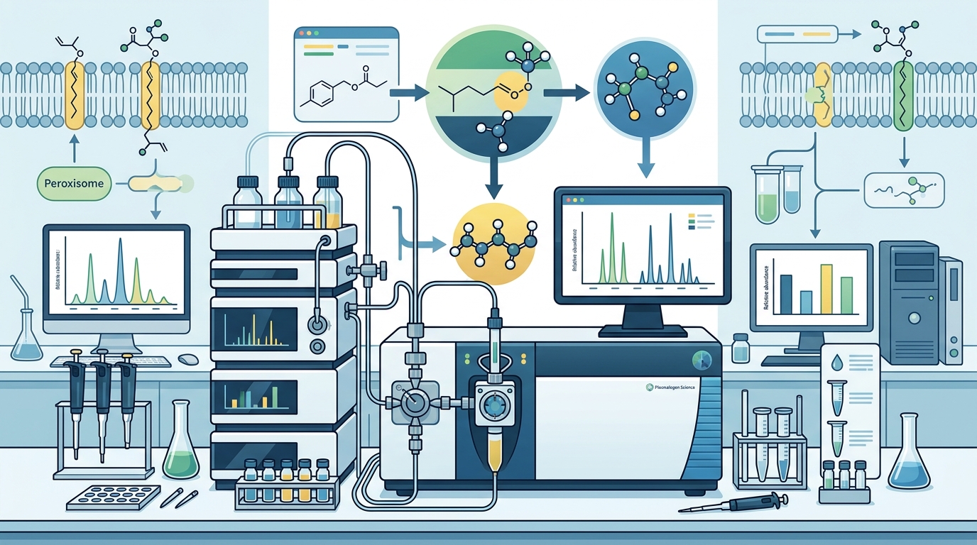
LC-MS/MS is the workhorse of modern plasmalogen analysis. In this approach, lipids are separated chromatographically and then fragmented inside the mass spectrometer, allowing identification of individual molecular species by their unique mass-to-charge patterns. Ultra-high-performance liquid chromatography coupled with quadrupole time-of-flight mass spectrometry (UHPLC-QTOF-MS) is widely used for global lipid profiling with particular emphasis on plasmalogen species. Some laboratories pair high-resolution accurate-mass (HRAM) Orbitrap instruments with triple-quadrupole systems to combine discovery-phase screening with targeted quantification of specific plasmalogen species.
2. Shotgun Lipidomics
Shotgun lipidomics bypasses chromatographic separation entirely. Instead, lipid extracts are directly infused into the mass spectrometer. Researchers have developed quantitative shotgun lipidomics approaches specifically for the study of plasmalogens in neuronal cells, using the resulting data alongside immunocytochemistry and neurotransmitter assays to characterize synaptic biology. While shotgun methods offer speed, they can struggle to resolve isobaric species — molecules with identical nominal masses but different structures.
3. Gas Chromatography–Mass Spectrometry (GC-MS)
GC-MS remains important for measuring total plasmalogen content. The Mayo Clinic Laboratories, for instance, use a GC-MS protocol for their clinical plasmalogen blood test. In their method, samples are mixed with internal standards, derivatized, extracted, dried under nitrogen, reconstituted, and then analyzed by GC-MS. This approach excels at quantifying the dimethyl acetal (DMA) derivatives released from the sn-1 vinyl ether bond, providing a reliable measure of total plasmalogen pools rather than individual molecular species.
4. Trapped Ion Mobility Spectrometry (TIMS)
A newer entrant, Trapped Ion Mobility Spectrometry coupled with mass spectrometry adds a gas-phase separation dimension based on molecular shape. A 2025 study published in Analytical Chemistry benchmarked TIMS for differentiating plasmalogens from other ether lipids, representing an important step toward fast, reliable characterization of these lipid species in biological samples. By separating ions based on their collisional cross-section, TIMS can resolve plasmenyl and plasmanyl species that co-elute chromatographically.
5. Thin-Layer Chromatography (TLC) with Downstream Analysis
Historically, phospholipids were first fractionated into classes via TLC before molecular species were analyzed by gas chromatography. Although largely superseded by LC-MS workflows for research, TLC-based fractionation remains a valid preparatory step in some clinical and reference laboratories.
Key Biomarkers and Molecular Species
Not all plasmalogens carry equal clinical weight. The specific species measured and the biological matrix sampled determine the biomarker's utility.
Ethanolamine Plasmalogens (PlsEtn)
PlsEtn species — particularly those esterified with docosahexaenoic acid (DHA, 22:6) at the sn-2 position — are the most extensively studied plasmalogen biomarkers. Decreased blood levels of ethanolamine plasmalogens and related lipids have been consistently observed in Alzheimer's disease across multiple independent studies. PlsEtn species are the predominant plasmalogen class in neural tissue, making them especially relevant for neurological assessment.
Choline Plasmalogens (PlsCho)
Choline plasmalogens are abundant in cardiac tissue and are also present in the brain. Postmortem lipidomic studies using UPLC-MS/MS have revealed significant decreases in choline plasmalogens containing DHA and stearic acid in the prefrontal cortex of Alzheimer's patients compared to healthy controls. PlsCho species are considered complementary biomarkers to PlsEtn in multi-system health assessment.
Red Blood Cell Plasmalogen Ratios
Clinical laboratories often measure plasmalogen content in erythrocyte membranes. The Mayo Clinic blood test specifically quantifies C16:0, C18:0, and C18:1 plasmalogens in red blood cells, normalized against C16:0 and C18:0 fatty acids. These ratios are the standard diagnostic tool for peroxisomal biogenesis disorders, including Zellweger syndrome spectrum and rhizomelic chondrodysplasia punctata (RCDP).
Oxidative Stress Co-Biomarkers
Because plasmalogens act as endogenous antioxidants — sacrificing their vinyl-ether bond to neutralize reactive oxygen species — clinicians often pair plasmalogen measurements with oxidative stress markers such as malondialdehyde (MDA), superoxide dismutase (SOD), and catalase. In a clinical trial of cognitively impaired persons receiving DHA-specific plasmalogen precursors, a dose-dependent elevation in DHA-plasmalogen levels correlated with improvements in these oxidative stress biomarkers.
DHA-Plasmalogen and Arachidonic Acid-Plasmalogen Subspecies
The fatty acid at sn-2 determines both function and clinical significance. DHA-containing plasmalogens are concentrated in synaptic membranes and retinal tissue, while arachidonic acid-containing species are enriched in immune and inflammatory cells. Subspecies-level profiling is becoming essential for distinguishing neurological from inflammatory or metabolic plasmalogen deficiency patterns.
Clinical Tests Available Today
Mayo Clinic Laboratories — Plasmalogens, Blood (PGRBC)
This established clinical test measures erythrocyte plasmalogens via GC-MS and is primarily ordered when diagnosing peroxisomal disorders. It quantifies C16:0, C18:0, and C18:1 species with fatty acid normalization. The test was developed in a manner consistent with CLIA requirements and serves as a diagnostic marker for conditions like Zellweger syndrome and RCDP.
Multi-System Plasmalogen Panels
Newer commercial panels, such as those offered by functional medicine laboratories, measure approximately 100 biomarkers organized into biosystem categories associated with advanced health and longevity. These panels go beyond simple plasmalogen quantification to contextualize results within broader metabolic, inflammatory, and lipid profiles — providing a multi-dimensional view of biochemical health.
Research-Grade Lipidomics Services
For academic and pharmaceutical research, specialized lipidomics service providers offer high-resolution LC-MS/MS plasmalogen profiling with GC-MS verification and redox-safe workflows designed to preserve vinyl-ether integrity during sample preparation. These services distinguish between P- and O- ether lipid species and provide detailed subspecies quantification across complex biological matrices.
Sample Handling and Pre-Analytical Considerations
Accurate plasmalogen measurement depends heavily on pre-analytical handling. The vinyl-ether bond is susceptible to oxidative degradation, which means poor sample handling can produce falsely low results.
- Temperature: Samples should be shipped on dry ice and stored at −80°C prior to extraction.
- Light protection: Amber vials or foil wrapping minimize photo-oxidation of the vinyl-ether bond.
- Extraction method: The Folch extraction method is commonly used for lipid isolation from tissue and blood samples before LC-MS analysis.
- Metadata: Including sample matrix type, species or strain, harvest time point, and treatment details supports accurate normalization and result interpretation.
- Antioxidant additives: Some protocols add butylated hydroxytoluene (BHT) to extraction solvents to prevent artifactual oxidation during processing.
Interpreting Your Plasmalogen Results
Interpreting plasmalogen biomarkers requires clinical context. A low result may reflect impaired peroxisomal biosynthesis, elevated oxidative stress consuming plasmalogens faster than they are produced, dietary insufficiency of precursor lipids, or age-related biosynthetic decline.
Context-Dependent Interpretation
| Finding | Possible Implication | Follow-Up Considerations |
|---|---|---|
| Low RBC C16:0/C18:0 plasmalogen ratios | Peroxisomal biogenesis disorder | Genetic testing for PEX genes, GNPAT, or AGPS |
| Low serum PlsEtn-DHA | Neurological risk, oxidative stress | Cognitive assessment, oxidative stress panel |
| Decreased PlsCho in cardiac context | Cardiac membrane vulnerability | Cardiovascular risk evaluation |
| Low plasmalogens with elevated MDA | Active oxidative consumption | Antioxidant status review, dietary intervention |
| Age-appropriate decline | Normal aging trajectory | Longitudinal monitoring, preventive strategies |
A single measurement provides a snapshot. Serial testing over months reveals whether interventions — dietary changes, supplementation with plasmalogen precursors, or lifestyle modifications — are shifting the trajectory in the right direction.
Disease Associations Revealed by Plasmalogen Biomarkers
Plasmalogen deficiency is not limited to rare genetic disorders. Altered plasmalogen metabolism has been described across a broad disease spectrum.
- Alzheimer's Disease: Multiple studies report that AD patients have lower levels of plasmalogen species, particularly those containing DHA, in both red blood cells and plasma compared to controls.
- Parkinson's Disease: Oral administration of ether phospholipids has been associated with improvement of blood plasmalogens and clinical symptoms in preliminary reports.
- Cancer: Lipidomics-based screening has identified plasmalogens as potential biomarkers for colon cancer, with plasma lipid profiles enabling discrimination between cancer patients and control subjects.
- Schizophrenia: Dysfunctional plasmalogen dynamics have been documented in both plasma and platelets of patients with schizophrenia.
- Peroxisomal Disorders: Severe plasmalogen deficiency is the hallmark biochemical finding in RCDP types 1, 2, and 3, as well as Zellweger syndrome spectrum disorders.
Key Takeaways
- Multiple platforms exist: LC-MS/MS, GC-MS, shotgun lipidomics, and emerging TIMS-based methods each serve different analytical purposes, from clinical diagnosis to deep research profiling.
- PlsEtn and PlsCho are core biomarkers: Ethanolamine and choline plasmalogens — especially DHA-containing subspecies — are the most clinically informative lipid fractions to track.
- Clinical testing is available now: Established tests like Mayo Clinic's PGRBC panel diagnose peroxisomal disorders, while newer multi-system panels assess plasmalogens as part of broader metabolic health optimization.
- Sample handling matters enormously: The vinyl-ether bond degrades under heat, light, and oxidative conditions, making pre-analytical care essential for accurate results.
- Context drives interpretation: Low plasmalogens can reflect genetic disorders, oxidative stress, dietary gaps, or normal aging — clinical history determines which pathway is most likely.
- Serial testing reveals trends: A single measurement is a snapshot; longitudinal tracking is the most valuable approach for assessing interventions and health trajectories.
Frequently Asked Questions
What is the most accurate method for measuring plasmalogen levels?
High-resolution LC-MS/MS is currently considered the gold standard for subspecies-level plasmalogen quantification. It can differentiate between plasmenyl (P-) and plasmanyl (O-) ether lipids and resolve individual molecular species by their unique fragmentation patterns. GC-MS remains reliable for measuring total plasmalogen content through dimethyl acetal derivatives.
Can I get a plasmalogen blood test from my doctor?
Yes. The Mayo Clinic Laboratories offer a clinical plasmalogen blood test (PGRBC) that measures erythrocyte plasmalogens via GC-MS. This test is primarily used for diagnosing peroxisomal disorders. Additionally, functional medicine laboratories now offer broader panels that include plasmalogen measurements alongside approximately 100 other biomarkers for health optimization purposes.
Which plasmalogen species are most important to track?
Ethanolamine plasmalogens containing DHA (PlsEtn-DHA) are the most frequently cited biomarkers in neurological research. Choline plasmalogens containing DHA and stearic acid (PlsCho) are additionally relevant for brain and cardiac health assessment. For peroxisomal disorder diagnosis, C16:0, C18:0, and C18:1 erythrocyte plasmalogens are the standard analytes.
Do plasmalogen levels decline with age?
Yes. Research consistently documents age-related shifts in ether lipid composition and metabolic flux. This decline is believed to reflect reduced peroxisomal biosynthetic capacity and accumulated oxidative damage over time. Longitudinal monitoring can help distinguish pathological decline from normal aging trajectories.
Why is sample handling so critical for plasmalogen testing?
The vinyl-ether bond that defines plasmalogens is highly susceptible to oxidative and photolytic degradation. If samples are exposed to heat, light, or air during collection, transport, or storage, the plasmalogens can break down before analysis, producing falsely low results. Proper protocols require dry-ice shipping, −80°C storage, amber or foil-wrapped vials, and sometimes the addition of antioxidants like BHT to extraction solvents.