
Plasmalogen Science traces the life cycle of plasmalogens—from the peroxisomal enzymes that build them to the synaptic membranes where they facilitate neurotransmission. This guide follows the molecule itself, showing how each stop along its journey supports a different aspect of brain health, membrane integrity, and myelin maintenance.
Where Plasmalogens Are Made: The Peroxisomal Starting Line
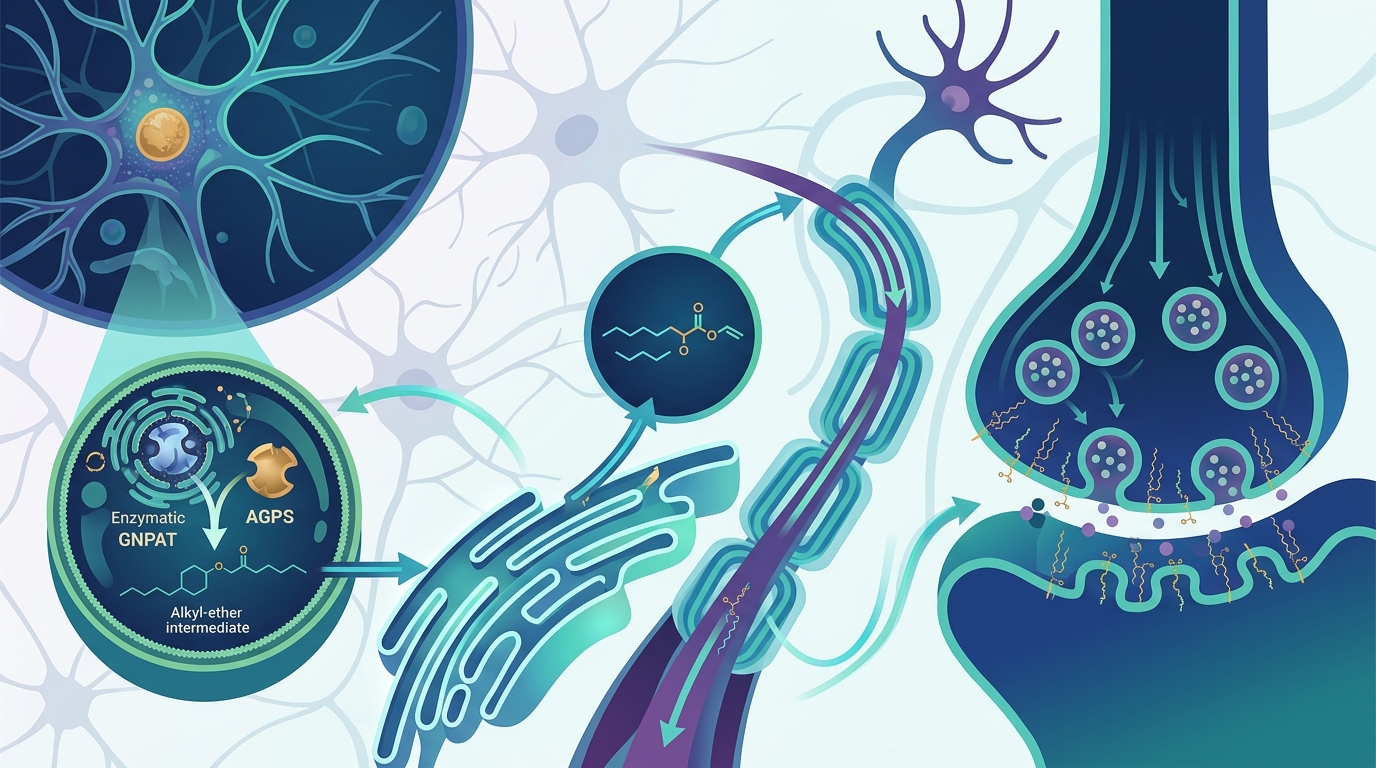
Every plasmalogen molecule begins inside a peroxisome, an organelle that most people associate with fatty-acid oxidation rather than lipid synthesis. Two peroxisomal enzymes—glyceronephosphate O-acyltransferase (GNPAT) and alkylglycerone phosphate synthase (AGPS)—catalyze the first two steps, generating the alkyl-ether intermediate that will later be converted to the signature vinyl-ether bond in the endoplasmic reticulum.
When these peroxisomal enzymes are absent, as in rhizomelic chondrodysplasia punctata (RCDP), the consequences are severe. Patients with RCDP present with myelination deficits, cerebellar atrophy, and enlarged ventricles—a clinical picture that underscores how essential the peroxisome-to-membrane pipeline is for the developing brain.
A Numbers Game: Plasmalogen Concentrations Across the Brain
Plasmalogens are not evenly distributed. They cluster in the tissues that demand the most structural support and the fastest signaling. Approximately one in every five phospholipids in human tissue is a plasmalogen, and the brain contains some of the highest concentrations of any organ.
Ethanolamine plasmalogen (PlsEtn) accounts for over 50 percent of total ethanolamine phosphoglycerides in gray matter and over 85 percent in myelin. Across the entire brain, plasmalogens constitute roughly 20 percent of total glycerophospholipids, while in myelin sheath ethanolamine glycerophospholipids, that figure climbs to as high as 70 percent.
Membrane Biophysics: How the Vinyl-Ether Bond Reshapes Lipid Bilayers
What sets plasmalogens apart from ordinary phospholipids is the vinyl-ether linkage at the sn-1 position of the glycerol backbone. This bond alters the geometry of the lipid in ways that propagate through the entire membrane.
Biophysical studies show that plasmalogens change the curvature, fluidity, rigidity, thickness, and lateral pressure of lipid bilayers. They also modulate the activity of integral membrane proteins by interacting directly with them. The glycerol backbone of plasmalogens frequently carries polyunsaturated fatty acids such as docosahexaenoic acid (DHA) and arachidonic acid (AA) at the sn-2 position, linking these lipids to both anti-inflammatory and pro-inflammatory signaling cascades.
For neurons, membrane fluidity is not an abstract property—it determines how efficiently ion channels open, how quickly receptors respond to ligands, and how readily synaptic vesicles merge with the presynaptic membrane.
Inside Myelin: Why White Matter Depends on Plasmalogens
Myelin is often described as the insulation around nerve fibers, but its composition matters as much as its presence. Plasmalogens are a fundamental structural component of the myelin sheath, and the highest concentration of plasmalogens in the entire body is found in white matter.
Within white matter, plasmalogen content directly influences nerve conduction velocity. In conditions like multiple sclerosis, where demyelination and chronic inflammation are hallmarks, studies show that low plasmalogen levels correlate with disease severity. Research also suggests that restoring plasmalogen levels may promote remyelination—the repair and regeneration of damaged myelin.
The connection between plasmalogens and Schwann cell differentiation in the peripheral nervous system further highlights the role these lipids play in myelination. A 2014 study published in the Journal of Clinical Investigation demonstrated that plasmalogens regulate Schwann cell differentiation and myelination in peripheral nerves.
At the Synapse: Vesicle Fusion and Signal Transmission
Plasmalogens have been described as "fusogenic"—they promote the merger of lipid bilayers. At the synapse, this property is critical. Neurotransmitter release depends on synaptic vesicles fusing with the presynaptic plasma membrane, and plasmalogens facilitate this process by introducing negative curvature stress into the membrane at fusion sites.
Animal studies have shown that plasmalogen supplementation in aged mice alleviates hippocampal synaptic loss and promotes both synaptogenesis and synaptic vesicle formation. In a 2022 study, two months of intragastric plasmalogen administration to aged C57BL/6J mice resulted in improved cognitive performance and measurable increases in synaptic density observed through transmission electron microscopy.
When plasmalogen levels fall, impaired vesicular fusion can compromise choline uptake at the nerve terminal, contributing to the cholinergic dysfunction that characterizes Alzheimer's disease.
The Sacrificial Antioxidant Mechanism
The vinyl-ether bond gives plasmalogens a unique antioxidant capability. Reactive oxygen species (ROS) preferentially attack this bond rather than the polyunsaturated fatty acids deeper in the membrane. In effect, plasmalogens function as sacrificial molecules, absorbing oxidative damage to protect neighboring lipids, proteins, and membrane structures.
This mechanism is especially important in the brain, which consumes roughly 20 percent of the body's oxygen despite comprising only about 2 percent of body weight. The high metabolic activity of neurons generates substantial ROS, making the antioxidant properties of plasmalogens an essential part of neural defense.
Plasmalogens also bolster broader antioxidant defenses. They modulate mitochondrial efficiency and support ROS scavenging pathways, creating a multi-layered protective system within neuronal membranes.
What Plasmalogen Deficiency Looks Like in the Brain
The clearest evidence for how critical plasmalogens are to the brain comes from studying what happens when they are absent or depleted.
Genetic Deficiency: RCDP
Rhizomelic chondrodysplasia punctata is caused by mutations in the PEX7 gene, which encodes a peroxisomal protein necessary for plasmalogen biosynthesis. Neurological abnormalities in RCDP include delayed myelination, enlarged ventricles, and cerebellar atrophy.
Alzheimer's Disease
Research using electrospray ionization mass spectrometry has revealed a dramatic decrease in plasmalogen content—up to 40 mol percent of total plasmalogen—in white matter at very early stages of Alzheimer's disease. In gray matter, the deficiency correlates with dementia severity, ranging from roughly 10 mol percent loss in very mild dementia to approximately 30 mol percent loss in severe dementia.
Plasmalogen levels decline with age broadly, but they decline more sharply and earlier in people who develop Alzheimer's disease and other forms of dementia. Research across five independent populations has found significant reductions in blood plasmalogens in dementia patients compared with controls, with the severity of depletion correlating with disease severity.
Multiple Sclerosis
People diagnosed with MS have significantly lower plasmalogen levels. This deficiency weakens nerve cell membranes, accelerates myelin degradation, and increases susceptibility to oxidative damage. Restoring plasmalogen levels may promote remyelination and support immune system balance in MS patients.
Parkinson's Disease
Plasmalogen levels are significantly reduced in Parkinson's disease patients. Research suggests that plasmalogens help protect dopaminergic neurons from oxidative stress and lipid peroxidation, and supplementation may support mitochondrial function and reduce neuroinflammation.
Age-Related Decline and Cognitive Consequences
Plasmalogen levels decrease as a natural part of aging, but the trajectory matters. A longitudinal study presented at the Alzheimer's Association International Conference found that a decrease in the plasmalogen index (the plasmalogen-to-phosphatidyl ratio) from baseline was associated with higher odds of converting from normal cognition to mild cognitive impairment or Alzheimer's disease (OR = 1.45, P = .002). Conversely, a higher plasmalogen index at baseline was associated with lower odds of conversion (OR = 0.63, P < .001).
Data from the Rush University Memory and Aging Project have revealed strong correlations between higher plasmalogen levels and reduced dementia risk, pointing to a key role for these lipids in cognitive longevity.
These findings raise an important question: is plasmalogen decline a cause or a consequence of neurodegeneration? The evidence increasingly supports a causal role, though the relationship is likely bidirectional—initial deficiencies accelerate pathology, which in turn depletes plasmalogens further.
Lessons from Animal Models
Recent animal research has strengthened the case for plasmalogen supplementation as a neuroprotective strategy.
- Aged mice fed plasmalogens for two months showed improved cognition and memory, reduced neuroinflammation, and evidence of both synaptogenesis and neurogenesis in hippocampal tissue.
- Pex7 knockout mice (which cannot produce plasmalogens) showed improved nerve conduction in peripheral nerves when treated with alkyl glycerol precursors before pathology onset, providing direct experimental evidence that restoring plasmalogen levels can ameliorate neurodegeneration.
- A novel inducible GNPAT knockout mouse model developed in 2024 allows researchers to induce plasmalogen deficiency in adult animals, mirroring the chronic decline observed in Alzheimer's disease. This model showed significant plasmalogen reductions in both circulation and tissues within four weeks of induction.
Key Takeaways
- Plasmalogens are built in peroxisomes and then distributed to membranes throughout the body, with especially high concentrations in the brain and myelin.
- The vinyl-ether bond is the defining structural feature, altering membrane geometry and providing a built-in antioxidant mechanism.
- Myelin depends heavily on plasmalogens—up to 70 percent of myelin ethanolamine glycerophospholipids are plasmalogens, and deficiencies are linked to demyelination.
- Synaptic function requires plasmalogen-driven membrane fusion, and depleted levels impair neurotransmitter release.
- Plasmalogen deficiency is a consistent finding in Alzheimer's disease, Parkinson's disease, and multiple sclerosis, with depletion severity correlating to disease progression.
- Age-related plasmalogen decline is a measurable risk factor for cognitive impairment, and higher baseline levels are protective.
- Animal studies show that plasmalogen supplementation can reverse synaptic loss, reduce neuroinflammation, and improve cognition in aged subjects.
Frequently Asked Questions
- What are plasmalogens?
- Plasmalogens are a class of phospholipids characterized by a vinyl-ether bond at the sn-1 position of the glycerol backbone. They make up roughly 20 percent of total phospholipids in human cell membranes and are concentrated in brain tissue, myelin, heart, and immune cells.
- Why are plasmalogens important for the brain?
- The brain relies on plasmalogens for membrane fluidity, synaptic vesicle fusion, antioxidant protection, and myelin sheath integrity. Their depletion is consistently associated with neurodegenerative diseases including Alzheimer's and Parkinson's.
- How do plasmalogens protect myelin?
- Plasmalogens constitute up to 70 percent of myelin ethanolamine glycerophospholipids. They maintain the structural integrity of the myelin sheath, protect it from oxidative damage, and support the differentiation of myelinating cells such as Schwann cells and oligodendrocytes.
- Do plasmalogen levels decline with age?
- Yes. Plasmalogen levels decrease as part of normal aging, but the decline is more pronounced in individuals who develop dementia. Longitudinal studies suggest that the rate and degree of decline can predict cognitive outcomes.
- What happens when the body cannot produce plasmalogens?
- Genetic disorders like rhizomelic chondrodysplasia punctata (RCDP), caused by mutations in peroxisomal genes, result in an inability to synthesize plasmalogens. Neurological consequences include delayed myelination, cerebellar atrophy, and severely impaired brain development.
- Can plasmalogen supplementation help brain function?
- Animal studies show promising results, including reduced neuroinflammation, improved synaptic density, and better cognitive performance in aged mice. Human clinical data remain limited, and questions about oral bioavailability and optimal dosing are still being investigated.