
Most discussions of dietary fats center on omega-3 fatty acids or cholesterol. Rarely do they mention the specialized phospholipids woven into every cell membrane you own—molecules that double as antioxidant shields, membrane architects, and signaling hubs. Those molecules are plasmalogens, and emerging research places them at the intersection of brain health, cardiovascular resilience, and the biology of aging itself.
This guide takes a different approach from typical overviews. Instead of organ-by-organ cataloging, it follows the life cycle of a plasmalogen molecule—from its birth inside peroxisomes, through the roles it performs in living membranes, to the consequences when production falters with age—so you can understand not just what plasmalogens do, but why their decline matters so profoundly.
Defining Plasmalogens: Chemistry in One Paragraph
Plasmalogens are a subclass of glycerophospholipids distinguished by a vinyl-ether bond at the sn-1 position of the glycerol backbone, instead of the more common ester linkage found in most phospholipids. An ester bond connects the sn-2 position to a fatty acyl chain—often a polyunsaturated fatty acid such as DHA or arachidonic acid. The head group is typically ethanolamine (forming plasmenylethanolamine, or PlsEtn) or choline (forming plasmenylcholine, or PlsCho). That single vinyl-ether bond is what gives plasmalogens their unique physical, chemical, and biological properties.
In quantitative terms, plasmalogens account for roughly 15–20 mol% of the total phospholipid mass in human tissues, though their abundance varies considerably by tissue type. Ethanolamine plasmalogens are the dominant form in most tissues, constituting approximately 5–20% of total phospholipids, while choline plasmalogens are concentrated primarily in cardiac and skeletal muscle membranes.
Birth of a Plasmalogen: The Peroxisome-to-ER Assembly Line
Unlike most phospholipids, plasmalogen synthesis requires two organelles working in tandem. The pathway begins inside peroxisomes and finishes at the endoplasmic reticulum (ER), proceeding through approximately seven enzymatic steps.
Step 1–2: Peroxisomal Initiation
Biosynthesis starts when the peroxisomal enzyme glyceronephosphate O-acyltransferase (GNPAT) acylates dihydroxyacetone phosphate (DHAP). Next, alkylglycerone phosphate synthase (AGPS) replaces the acyl group with a long-chain fatty alcohol, producing 1-O-alkyl-DHAP—the first ether-linked intermediate. GNPAT and AGPS physically associate on the luminal side of the peroxisomal membrane, increasing catalytic efficiency.
The FAR1 Bottleneck
The fatty alcohols consumed by AGPS are generated by fatty acyl-CoA reductase 1 (FAR1), anchored on the cytoplasmic face of the peroxisome. FAR1 is the rate-limiting enzyme in the pathway: the cell regulates plasmalogen output by sensing plasmalogen concentration in the inner leaflet of the plasma membrane and adjusting FAR1 protein stability accordingly. When plasmalogen levels are high, FAR1 is degraded faster; when levels fall, FAR1 is stabilized and production ramps up. This elegant feedback loop keeps plasmalogen homeostasis in check—until aging or disease overwhelms it.
Steps 3–7: ER Completion
The alkyl-DHAP intermediate exits the peroxisome and enters the ER, where it undergoes reduction, acylation at sn-2, head-group attachment, and finally desaturation by plasmanylethanolamine desaturase (PEDS1/TMEM189)—the enzyme that introduces the defining vinyl-ether double bond. Discovery of PEDS1 as the key desaturase was a landmark achievement, emerging from studies on how the myxobacterium Myxococcus xanthus responds to light.
Why Peroxisomes Matter
Because the first two steps are peroxisome-exclusive, any defect in peroxisome assembly cripples plasmalogen production. This principle is dramatically illustrated by Rhizomelic Chondrodysplasia Punctata (RCDP), a rare genetic disorder caused by mutations in GNPAT, AGPS, or the peroxisomal import factor PEX7. Patients with RCDP exhibit severely reduced plasmalogen levels and present with skeletal malformations, cataracts, seizures, impaired respiration, and abnormal neurological development—a clinical picture that reveals how many organ systems depend on adequate plasmalogen supply.
Life Inside the Membrane: Four Jobs No Other Lipid Can Do
Once inserted into the phospholipid bilayer, plasmalogens perform roles that ester-linked phospholipids cannot fully replicate.
1. Membrane Architecture and Fluidity
The vinyl-ether bond creates a more compact molecular conformation at the sn-1 position, packing adjacent phospholipids more tightly at the membrane-water interface. This influences bilayer thickness, curvature, and the formation of lipid raft microdomains—cholesterol-rich regions where signaling receptors cluster. Plasmalogens are important for the organization and stability of these rafts, directly affecting signal transduction efficiency.
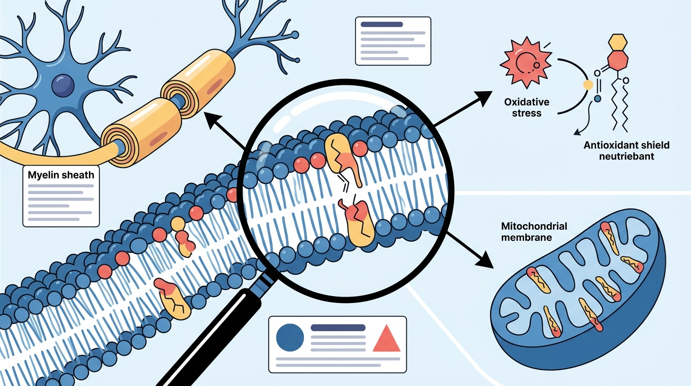
2. Endogenous Antioxidant Shield
The vinyl-ether bond is preferentially attacked by reactive oxygen species (ROS), making plasmalogens act as sacrificial antioxidants. When a ROS molecule cleaves the vinyl-ether bond, the plasmalogen is consumed but the adjacent polyunsaturated fatty acids and membrane proteins are spared. This is not a minor detail—it is a frontline defense mechanism embedded in every cell membrane.
3. PUFA Reservoir and Lipid Mediator Precursor
The sn-2 position of plasmalogens is enriched in polyunsaturated fatty acids, particularly DHA and arachidonic acid. This makes plasmalogens a reservoir for the release of these PUFAs, which serve as precursors to prostaglandins, thromboxanes, and other eicosanoid signaling molecules that regulate inflammation, blood clotting, and tissue repair.
4. Vesicular Fusion and Membrane Dynamics
Plasmalogens promote membrane curvature changes required for vesicle budding, fusion, and exocytosis. In synaptic transmission, where neurotransmitter vesicles must fuse rapidly with the presynaptic membrane, this property is indispensable.
Plasmalogens and the Brain: Myelin, Synapses, and Cognition
The human brain is among the most plasmalogen-rich organs. Ethanolamine plasmalogens are major components of both white and grey matter, though each compartment favors different molecular species. White matter plasmalogens tend to carry more saturated and monounsaturated side chains, consistent with their role in myelin—the insulating sheath around axons that enables rapid nerve conduction. Grey matter plasmalogens are enriched in DHA (22:6) and arachidonic acid (20:4), reflecting roles in synaptic membrane fluidity and signaling.
Disruption of plasmalogen homeostasis impairs cholesterol synthesis in the brain and reduces the expression of myelin basic protein, as demonstrated in plasmalogen-deficient mouse models. This connection between plasmalogen levels and myelination helps explain why peroxisomal disorders with low plasmalogens invariably feature white matter abnormalities.
Animal studies published in 2025 added further evidence: in a mouse model of age-related cognitive decline, plasmalogen supplementation improved spatial memory by approximately 44% and boosted the synaptic scaffolding protein PSD-95 in the hippocampus while reducing markers of brain inflammation. Notably, plasmalogens outperformed other phospholipids—including phosphatidylcholine and phosphatidylserine—across cognitive and biochemical measures.
The Age-Related Decline: What the Numbers Show
Plasmalogen levels do not remain constant across the human lifespan. Research indicates that brain plasmalogen concentrations decrease by roughly 40% between age 40 and age 70. Multiple factors drive this decline: reduced synthesis of DHA and its incorporation into ethanolamine plasmalogens, increased activity of the enzyme that degrades ethanolamine plasmalogens (PlsEtn-specific phospholipase A₂), chronic oxidative stress that consumes the vinyl-ether bond faster than cells can replace it, and mitochondrial dysfunction that further destabilizes plasmalogen metabolism.
A key insight from population studies is that the decline may not be uniform. Analysis of serum PlsEtn 16:0/22:6 levels across age decades revealed that a significant subpopulation experiences a dramatic drop between ages 50 and 69, while another subpopulation shows relatively little decline. This heterogeneity suggests that plasmalogen loss is not simply an inevitable consequence of calendar age but may be influenced by genetics, lifestyle, oxidative burden, and peroxisomal health.
Alzheimer's Disease and Plasmalogen Deficiency
The association between low plasmalogens and Alzheimer's disease (AD) is one of the most studied aspects of plasmalogen biology. Reductions of PlsEtn levels have been reported in plasma, serum, cerebrospinal fluid, and post-mortem brain tissue of AD patients. Some researchers have estimated that plasmalogens begin to decrease up to seven years before clinical diagnosis of dementia.
In the Alzheimer's Disease Neuroimaging Initiative (ADNI) cohort of 1,547 subjects, statistically significant negative relationships were found between AD diagnosis and plasmalogen-derived indices such as PLPX and the plasmalogen biosynthesis value (PBV). Aging-related declines in circulating plasmalogens containing omega-3 fatty acids may increase AD risk by reducing plasmalogen availability to the central nervous system.
A separate study tracking 40 AD patients over one year found that ADAS-Cog cognitive scores worsened significantly only in patients whose baseline serum plasmalogen levels were ≤75% of age-matched controls. Patients with normal plasmalogen levels showed no cognitive decline during the same period. While this was a pilot study, the pattern was consistent across mild, moderate, and severe plasmalogen depletion groups.
Whether plasmalogen deficiency is a cause, a consequence, or an accelerator of AD pathology remains an open question. The evidence supports all three possibilities to varying degrees, and many researchers now consider PlsEtn species as candidate AD biomarkers warranting further investigation.
Measuring Plasmalogen Status: Lipidomics and Biomarkers
Advances in mass spectrometry–based lipidomics have made it feasible to quantify individual plasmalogen species from blood samples. Key approaches include:
- Serum or plasma PlsEtn panels: Targeted assays measure specific ethanolamine plasmalogen species (e.g., PlsEtn 16:0/22:6, PlsEtn 18:0/20:4) that correlate with tissue levels and disease states.
- Composite indices: Researchers have developed aggregate scores—such as the Plasmalogen Biosynthesis Value (PBV) and PlsEtn/PtdEtn ratios—that capture the biosynthetic capacity of the pathway rather than just the concentration of a single species.
- Erythrocyte membrane analysis: Red blood cell membranes offer a window into longer-term plasmalogen status because erythrocytes lack nuclei and cannot synthesize new plasmalogens, making their membrane composition a time-averaged indicator.
These tools are moving from research laboratories toward clinical application. Plasmalogen profiling has been proposed as a surrogate marker of systemic oxidative stress, and reduced circulating plasmalogens have been associated with increased cardiovascular mortality in patients on renal replacement therapy.
Strategies for Supporting Plasmalogen Levels
Because plasmalogens sit at the crossroads of peroxisomal function, antioxidant defense, and membrane integrity, strategies to maintain their levels touch multiple biological systems.
Dietary Sources
Plasmalogens are found in animal-derived foods, particularly organ meats, shellfish, and marine invertebrates such as sea squirts (ascidians) and scallops. These organisms are rich in ethanolamine plasmalogens with DHA at the sn-2 position—the exact species most associated with brain and heart benefits.
Precursor Supplementation
Some research groups have explored orally administered plasmalogen precursors—alkylglycerol compounds designed to bypass the peroxisomal bottleneck. In a small clinical study, oral administration of a plasmalogen precursor increased serum plasmalogen levels, reduced signs of oxidative stress, and showed preliminary evidence of cognitive benefit in cognitively impaired participants.
Lifestyle and Metabolic Factors
Because peroxisomes are central to biosynthesis, any factor that supports peroxisomal biogenesis and function may indirectly support plasmalogen levels. Regular aerobic exercise has been linked to improved peroxisomal activity. Reducing chronic oxidative stress through balanced nutrition, adequate sleep, and avoidance of environmental toxins helps slow the rate at which plasmalogens are consumed as sacrificial antioxidants. Maintaining mitochondrial health is also relevant, since mitochondrial dysfunction accelerates plasmalogen degradation.
Note: Plasmalogen research is advancing rapidly, but most human intervention studies remain small or preliminary. Always consult a qualified healthcare provider before making supplementation decisions.
Key Takeaways
- Plasmalogens are unique phospholipids defined by a vinyl-ether bond at the sn-1 position, making up 15–20 mol% of membrane phospholipids in human tissues.
- Biosynthesis starts in peroxisomes and finishes in the endoplasmic reticulum, with FAR1 acting as the rate-limiting checkpoint regulated by a membrane-sensing feedback loop.
- Four irreplaceable membrane functions: structural organization of lipid rafts, sacrificial antioxidant protection, PUFA reservoir for signaling lipids, and facilitation of vesicular fusion.
- The brain is especially dependent on plasmalogens for myelin integrity, synaptic function, and cholesterol metabolism.
- Levels decline with age—brain concentrations may drop ~40% between ages 40 and 70—driven by reduced synthesis, increased degradation, and oxidative stress.
- Low plasmalogens correlate with Alzheimer's disease, and the decline may precede clinical diagnosis by up to seven years.
- Lipidomics now enables measurement of individual plasmalogen species and composite biosynthesis indices from standard blood samples.
- Dietary plasmalogens, precursor supplements, and lifestyle factors that support peroxisomal and mitochondrial health may help preserve levels, though large-scale clinical trials are still needed.
Frequently Asked Questions
What exactly are plasmalogens?
Plasmalogens are a subclass of glycerophospholipids characterized by a vinyl-ether bond at the sn-1 position of the glycerol backbone. They are found in virtually every human cell membrane and are especially concentrated in the brain, heart, kidneys, lungs, and skeletal muscle. Their two major forms are ethanolamine plasmalogens (PlsEtn) and choline plasmalogens (PlsCho).
Why are plasmalogens important for brain health?
Plasmalogens are critical structural components of myelin sheaths and synaptic membranes. They support cholesterol metabolism in the brain, serve as reservoirs for DHA, and protect neural membranes from oxidative damage. Research shows that reduced brain plasmalogen levels are associated with impaired cognition and neurodegenerative diseases such as Alzheimer's.
Do plasmalogen levels decrease with age?
Yes. Studies indicate that brain plasmalogen concentrations can decline by approximately 40% between ages 40 and 70. This decline results from reduced biosynthesis, increased enzymatic degradation, chronic oxidative stress, and mitochondrial dysfunction. However, the rate of decline varies between individuals.
How are plasmalogens synthesized in the body?
Plasmalogen biosynthesis begins in peroxisomes, where the enzymes GNPAT and AGPS create the initial ether-linked intermediate. The process then moves to the endoplasmic reticulum for completion across approximately seven enzymatic steps. FAR1, the enzyme that produces fatty alcohols for the pathway, serves as the rate-limiting step and is regulated by a feedback mechanism that senses membrane plasmalogen levels.
Can you increase plasmalogen levels through diet?
Plasmalogens are present in animal-derived foods such as organ meats, shellfish, scallops, and sea squirts. Some clinical studies have explored plasmalogen-enriched supplements and oral precursors that may raise circulating plasmalogen levels. Research is still in early stages, and you should consult a healthcare professional before beginning any supplementation protocol.
What is the connection between plasmalogens and Alzheimer's disease?
Multiple studies have found that ethanolamine plasmalogen levels are significantly reduced in the blood, cerebrospinal fluid, and brain tissue of Alzheimer's patients. Some evidence suggests that this decline begins years before clinical symptoms appear. Whether low plasmalogens are a cause, a consequence, or an accelerating factor in Alzheimer's pathology is still being investigated, but PlsEtn species are increasingly considered as candidate biomarkers for the disease.