
Most conversations about healthy fats stop at omega-3s. But buried inside every cell membrane sits a class of phospholipids that science is only now placing at center stage: plasmalogens. From insulating neurons to shielding cardiac muscle from oxidative attack, plasmalogens serve functions no other lipid can replicate. This guide maps their influence across seven organ systems, explains how your body makes them, and reviews what happens when levels fall.
The Vinyl-Ether Bond: What Sets Plasmalogens Apart
All phospholipids share a glycerol backbone, but plasmalogens differ at the sn-1 position, where a vinyl-ether bond replaces the usual ester linkage. That single structural quirk gives the molecule unique physical and chemical properties. Plasmalogens are a unique family of cellular glycerophospholipids defined by this vinyl-ether bond, and they account for roughly 15–20 mol% of total phospholipid mass in mammalian tissues.
Two major classes dominate human biology:
- Ethanolamine plasmalogens (PlsEtn) — enriched in brain and nervous tissue
- Choline plasmalogens (PlsCho) — concentrated in heart muscle
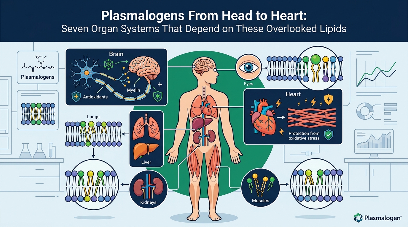
Brain myelin possesses the highest content of ethanolamine plasmalogens, whereas heart muscle carries a higher proportion of choline plasmalogens. Moderate amounts appear in kidney, skeletal muscle, spleen, and blood cells, while the liver maintains comparatively low levels.
The vinyl-ether bond is reactive toward oxidants, which makes plasmalogens inherently unstable under oxidative or inflammatory stress. Far from a design flaw, this reactivity is the basis of their role as sacrificial antioxidants: the bond intercepts reactive oxygen species before they can damage adjacent polyunsaturated fatty acid chains or membrane proteins.
Peroxisomal Origins: How Your Cells Build Plasmalogens
Unlike most phospholipids, plasmalogen assembly begins inside peroxisomes—small, single-membrane organelles best known for fatty-acid oxidation. Plasmalogen biosynthesis is initiated in the peroxisome by the enzymes glyceronephosphate O-acyltransferase (GNPAT) and alkylglycerone phosphate synthase (AGPS) and is then completed in the endoplasmic reticulum.
A built-in feedback loop keeps supply matched to demand. Plasmalogen biosynthesis is spatiotemporally regulated by a mechanism that senses plasmalogen concentration in the inner leaflet of the plasma membrane and adjusts the stability of fatty acyl-CoA reductase 1 (FAR1), the rate-limiting enzyme in the pathway.
When peroxisomal function fails—as in genetic disorders like Zellweger syndrome or Rhizomelic Chondrodysplasia Punctata (RCDP)—plasmalogen levels collapse across multiple organs, producing severe neurological, skeletal, and respiratory symptoms.
System 1 — Brain and Cognitive Function
Plasmalogens are disproportionately concentrated in the central nervous system, where they contribute to membrane fluidity, vesicle fusion, and synaptic signaling. Emerging evidence indicates that plasmalogens may be beneficial for Alzheimer's disease and cognitive aging.
Research using non-targeted metabolomics observed consistent depletion of plasmalogens in individuals with cognitive impairment and Alzheimer's disease. One landmark study found a dramatic decrease in plasmalogen content—up to 40 mol% of total plasmalogen—in white matter at a very early stage of Alzheimer's disease (CDR 0.5, or very mild dementia). Gray-matter deficiency correlated with disease severity, ranging from approximately 10 mol% at CDR 0.5 to roughly 30 mol% at CDR 3 (severe dementia).
Population-level data reinforce these findings. One analysis showed that an 85-year-old with high plasmalogen levels had only a 5% probability of having dementia, compared with 28% for an 85-year-old with low levels. Strikingly, a 95-year-old with high plasmalogen levels had the same dementia likelihood as a 75-year-old with low levels, suggesting that robust plasmalogen status may effectively subtract 20 years of cognitive-aging risk.
System 2 — Myelin and White Matter
Myelin—the insulating sheath that enables rapid nerve conduction—is arguably the tissue most dependent on plasmalogens. Plasmalogens are the most abundant class of phospholipids in myelin, making up some 80–90% of myelin membrane phospholipids.
Deficiency of plasmalogens causes profound abnormalities in the myelination of nerve cells, which is one reason why many peroxisomal disorders lead to neurological disease. Plasmalogen-knockout mice develop defects in central nervous system myelination, cataracts, and arrest of spermatogenesis. Furthermore, dysregulation of plasmalogen synthesis impairs cholesterol synthesis in the brain, resulting in reduced expression of myelin basic protein—a phenotype observed in the cerebellum of plasmalogen-deficient mice.
System 3 — Cardiovascular Tissue
The heart is one of the most plasmalogen-rich organs in the body. Plasmalogens are enriched in cardiac tissue and positively affect the cardiovascular system through antioxidant activity, support of healthy lipid profiles, and reduction of atherosclerosis risk.
The Heart SCORE study—a community-based investigation following 1,852 participants for approximately 16 years—found that mid-life levels of plasmalogens and other anti-inflammatory metabolites were inversely associated with late-life atherosclerotic cardiovascular disease events. Separately, a higher Plasmalogen Score has been linked to reduced risk of type 2 diabetes, cardiovascular disease, and lower overall mortality.
In models of myocardial infarction, researchers identified chlorolipids—oxidation products formed when myeloperoxidase attacks the vinyl-ether bond—in damaged heart tissue, suggesting that plasmalogen consumption during cardiac events may itself contribute to tissue injury when reserves run low.
System 4 — Immune Cells and Inflammation
Plasmalogens are embedded throughout immune cell membranes and participate in inflammatory signaling at multiple levels. During inflammation, neutrophil-derived myeloperoxidase produces hypochlorous acid, which causes oxidative chlorination of plasmalogens at the sn-1 chain by reacting with the vinyl-ether bond. The resulting chlorolipids can themselves act as signaling molecules.
Additionally, plasmalogens interact with platelet-activating factor (PAF) pathways. Human studies have identified elevated PAF levels in patients with coronary artery disease, suggesting a role for plasmalogens in PAF interconversion or receptor antagonism. Altered plasmalogen metabolism has been described across a broad range of systemic and chronic diseases, from COVID-19 and sepsis to systemic lupus erythematosus.
System 5 — Lungs and Respiratory Membranes
Peroxisomes play a role in the biosynthesis of ether phospholipids critical for the normal function of mammalian brains and lungs. Pulmonary surfactant—the mixture that keeps alveoli from collapsing—contains significant amounts of plasmalogen species. Individuals with severe plasmalogen deficiencies frequently show impaired respiration alongside neurological and skeletal abnormalities.
System 6 — Eyes and Lens Integrity
Cataracts are a consistent finding in severe plasmalogen-deficiency disorders. Plasmalogen-knockout mice develop cataracts, highlighting the molecule's role in maintaining lens transparency. The lens is metabolically unique—cells in its interior lack organelles—so membrane lipid composition is critical to optical clarity over a lifetime.
System 7 — Reproductive Biology
Plasmalogen-knockout mice exhibit arrest of spermatogenesis, and disruption of the blood-testis barrier has been documented in ether-lipid-deficient models. While human reproductive studies are fewer, these animal data point to plasmalogens as structural requirements for normal gamete development and testicular compartmentalization.
Why Plasmalogen Levels Decline With Age
In healthy adults, plasmalogen levels peak in early adulthood and then decline slowly—roughly 40% may be lost by age 70. Multiple factors converge to drive this decline:
- Peroxisomal aging — Peroxisome number and enzymatic efficiency decrease with age, reducing biosynthetic capacity.
- Cumulative oxidative stress — Chronic exposure to reactive oxygen species consumes plasmalogens faster than aging peroxisomes can replace them.
- Chronic inflammation — Sustained immune activation accelerates vinyl-ether oxidation via myeloperoxidase pathways.
- Mitochondrial dysfunction — As mitochondrial efficiency falls, increased electron leakage generates more ROS, further depleting plasmalogens.
The oxidative susceptibility of plasmalogens renders them particularly vulnerable under inflammatory or oxidative stress, contributing to a measurable reduction in total plasmalogen content that tracks with disease progression.
Plasmalogen-deficient cells show increased membrane lipid mobility on fluorescence anisotropy assays and are more sensitive to ROS and cell death than wild-type cells—a reminder that depletion does not merely correlate with disease but may actively facilitate it.
Measuring Plasmalogens: Lipidomics and Biomarker Panels
A reduction of circulating plasmalogens is increasingly used clinically as a biomarker of disease. Modern lipidomic platforms—particularly tandem mass spectrometry (LC-MS/MS)—can resolve individual plasmalogen species by head group, chain length, and degree of unsaturation, providing far more granular data than earlier thin-layer chromatography methods.
Commercially available blood panels now measure key plasmalogen species, including DHA- and choline-based forms, as well as related lipid biomarkers. These profiles can identify patterns linked to higher risk of Alzheimer's disease, Parkinson's disease, and other neurodegenerative conditions. Clinical trials have also demonstrated that targeted plasmalogen precursor supplementation can raise blood plasmalogen levels and alter oxidative stress biomarkers in cognitively impaired persons.
Key Takeaways
- Plasmalogens are vinyl-ether phospholipids comprising 15–20% of mammalian membrane phospholipids, with unique antioxidant and structural properties.
- Their biosynthesis begins in peroxisomes and is completed in the endoplasmic reticulum; peroxisomal health is therefore a bottleneck for plasmalogen supply.
- Myelin is the most plasmalogen-dependent tissue, with 80–90% of its phospholipids belonging to this class.
- Depleted plasmalogen levels have been documented in Alzheimer's disease, cardiovascular disease, diabetes, and multiple systemic inflammatory conditions.
- Levels decline roughly 40% by age 70, driven by oxidative stress, inflammation, and peroxisomal aging.
- Blood-based lipidomic panels now allow clinicians to quantify plasmalogen status and assess disease risk.
- Higher plasmalogen levels in older adults are associated with dramatically lower dementia probability.
Frequently Asked Questions
What exactly are plasmalogens?
Plasmalogens are a subclass of glycerophospholipids distinguished by a vinyl-ether bond at the sn-1 position of the glycerol backbone. They represent approximately 15–20 mol% of total phospholipids in mammalian tissues and are especially concentrated in brain, heart, and immune cells. Their reactive vinyl-ether bond allows them to act as sacrificial antioxidants, neutralizing reactive oxygen species before they damage other membrane components.
Why do plasmalogen levels drop with age?
Plasmalogen levels decline because peroxisomal biosynthetic capacity diminishes over time while cumulative oxidative stress and chronic inflammation consume existing plasmalogen stores faster than they can be replaced. In healthy adults, levels may fall by roughly 40% by age 70.
How are plasmalogens connected to Alzheimer's disease?
Studies show up to 40 mol% plasmalogen depletion in brain white matter at the earliest clinical stage of Alzheimer's disease. Population data also indicate that individuals with high blood plasmalogen levels have a dramatically lower probability of developing dementia compared to those with low levels, even at advanced ages.
Do plasmalogens affect heart health?
Yes. The heart is one of the most plasmalogen-rich organs. Research from the Heart SCORE study found that higher mid-life plasmalogen levels were inversely associated with late-life cardiovascular events over roughly 16 years of follow-up. Plasmalogens support healthy lipid profiles and may reduce atherosclerosis risk through their antioxidant and anti-inflammatory properties.
Can plasmalogen levels be measured?
Yes. Blood-based lipidomic panels using liquid chromatography–tandem mass spectrometry (LC-MS/MS) can quantify individual plasmalogen species. Commercially available tests now measure key species, including DHA- and choline-based plasmalogens, and can identify patterns associated with heightened risk of neurodegenerative and cardiometabolic disease.
What role do peroxisomes play in plasmalogen production?
Peroxisomes initiate plasmalogen biosynthesis through the enzymes GNPAT and AGPS. The process is then completed in the endoplasmic reticulum. When peroxisomal function is impaired—as in Zellweger syndrome—plasmalogen levels collapse across multiple organs, causing severe neurological, skeletal, and respiratory dysfunction.