
Plasmalogens are among the most important — and most overlooked — lipids in your body. Found in high concentrations in the brain, heart, and immune cells, these vinyl-ether phospholipids are essential for membrane integrity, antioxidant defense, and cellular signaling. But how do researchers and clinicians actually measure plasmalogen levels, and what do the results mean for your health?
This guide walks through every major measurement method, the key biomarkers scientists track, the clinical tests currently available, and the disease associations that make plasmalogen testing increasingly relevant.
Why Measure Plasmalogen Levels?
Plasmalogens are not just structural fillers in cell membranes. Their biosynthesis begins in peroxisomes, making circulating plasmalogen concentrations a functional readout of peroxisomal health. As research from ScienceDirect has documented, plasma plasmalogens may reflect the systemic functional activity of peroxisomes and serve as potential biomarkers for diseases related to oxidative stress and aging.
Several lines of evidence make plasmalogen measurement clinically meaningful:
- Neurodegeneration: Reduced ethanolamine plasmalogen (PlsEtn) levels appear in the brain and serum of Alzheimer's disease patients, often years before clinical diagnosis.
- Peroxisomal disorders: Conditions such as Zellweger spectrum disorder (ZSD) and rhizomelic chondrodysplasia punctata (RCDP) are characterized by severely diminished plasmalogens.
- Aging: Age-related shifts in ether lipid composition are well documented in population-level lipidomics studies.
- Cancer: Altered plasmalogen profiles have been identified as potential biomarkers for differentiating colon cancer patients from healthy controls.
The Analytical Challenge of Plasmalogens
Measuring plasmalogens is not straightforward. Their defining feature — the vinyl-ether bond at the sn-1 position — is chemically labile and structurally similar to alkyl-ether (plasmanyl) lipids, creating identification problems. The presence of another isomeric ether-containing subclass of lipids in phosphatidylethanolamine (PE) and phosphatidylcholine (PC) often complicates the identification of plasmalogens in a cellular lipid mixture.
Additionally, plasmalogens are sensitive to oxidation, acid hydrolysis, and light exposure, meaning that pre-analytical conditions heavily influence results. These challenges have driven the development of increasingly sophisticated analytical platforms.
Major Measurement Methods
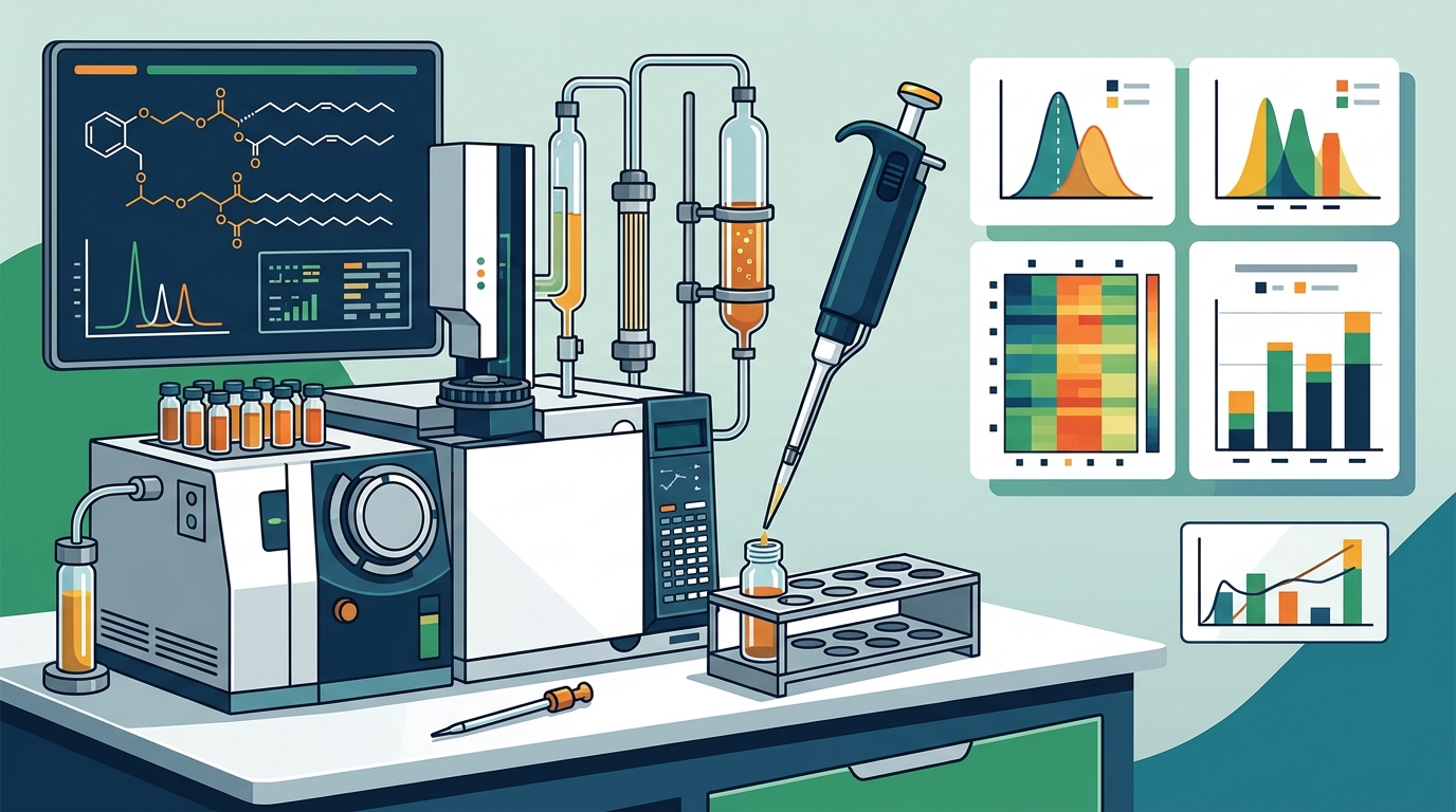
1. Gas Chromatography–Mass Spectrometry (GC-MS)
GC-MS has been the traditional clinical workhorse for plasmalogen analysis. The approach exploits the acid-labile vinyl-ether bond: after acid hydrolysis, plasmalogens release fatty aldehydes that are converted to dimethyl acetals (DMAs). These DMAs are then separated and quantified via GC-MS. This method can confirm true plasmalogens and resolve isobaric O-ether interferences.
The Mayo Clinic Laboratories uses a GC-MS-based assay that measures C16:0, C18:0, and C18:1 plasmalogens in red blood cells along with corresponding fatty acids for ratio normalization. This remains a standard clinical reference method for diagnosing peroxisomal disorders.
2. Liquid Chromatography–Tandem Mass Spectrometry (LC-MS/MS)
LC-MS/MS represents the current gold standard for detailed, structure-specific plasmalogen quantitation. Unlike GC-MS, LC-MS/MS can preserve the intact phospholipid molecule, allowing researchers to determine the specific fatty acid at the sn-2 position (commonly DHA or arachidonic acid) without hydrolysis.
Multiple reaction monitoring (MRM) transitions can be optimized to distinguish between PC plasmalogens and PE plasmalogens, quantifying individual molecular species like PlsEtn 16:0/22:6 or PlsEtn 18:0/20:4. Recent work has demonstrated LC-MS/MS as a viable clinical replacement for the older GC-MS method in diagnosing peroxisomal biogenesis disorders, offering improved structural specificity.
3. Shotgun Lipidomics (MDMS-SL)
Multi-dimensional mass spectrometry-based shotgun lipidomics (MDMS-SL) takes a different approach by analyzing lipid extracts directly without chromatographic separation. This platform enables the analysis of nearly 50 lipid classes and thousands of individual lipid species with high accuracy. It is particularly powerful for research-scale population studies but is less commonly used in clinical diagnostics.
4. Hydrophilic Interaction Chromatography–MS (HILIC-MS)
HILIC-MS is commonly used because of its sensitivity, resolution, and reproducibility for separation and detection of phospholipids. This method is especially valuable for characterizing the molecular species profile of plasmalogens, including the specific combination of head group, sn-1 fatty alcohol, and sn-2 fatty acid in each molecule.
5. Nuclear Magnetic Resonance (NMR) Spectroscopy
NMR provides a non-destructive approach to plasmalogen analysis that can identify the vinyl-ether bond directly through its distinctive chemical shift. While less sensitive than mass spectrometry, NMR is useful for confirming the structural identity of plasmalogens in research contexts.
6. Enzymatic Assay
Enzymatic assays offer a simpler, more scalable alternative to instrumental methods. These assays use enzymes that specifically act on the vinyl-ether bond or the phospholipid backbone to generate measurable products. Researchers have validated enzymatic assays alongside LC-MS/MS for plasma and serum plasmalogens, making them attractive for large-cohort studies.
7. ELISA-Based Methods
Enzyme-linked immunosorbent assay (ELISA) has been used in the research literature as one of three traditional methods for assessing plasmalogen levels in blood, alongside mass spectrometry and NMR. ELISA is antibody-based and can be adapted for high-throughput screening, though it typically offers less molecular detail than mass spectrometry.
Key Plasmalogen Biomarkers and Indices
Measuring total plasmalogens is useful, but modern lipidomics has identified several specific biomarkers and composite indices that carry distinct clinical meaning.
Ethanolamine Plasmalogens (PlsEtn)
PlsEtns are the most abundant plasmalogens in the brain and the most widely studied class in neurodegeneration research. Reductions of PlsEtn levels have been reported in plasma, serum, cerebrospinal fluid, and brain tissue of Alzheimer's disease patients. The specific molecular species PlsEtn 16:0/22:6 (containing DHA at sn-2) is frequently cited as a neurological biomarker of particular interest.
PlsEtn 16:0/22:6 (DHA Plasmalogen)
This individual species has emerged as a sentinel marker. Research published in the Journal of Lipid Research showed that a significant subpopulation exhibited a dramatic decline in serum PlsEtn 16:0/22:6 between the ages of 50 and 69, while a second subpopulation showed little decline with age — suggesting that plasmalogen decline is not merely a universal aging phenomenon but may identify individuals at elevated disease risk.
Plasmalogen Biosynthesis Value (PBV)
PBV is a composite index reflecting the overall capacity of peroxisomal plasmalogen synthesis. Studies using data from the Alzheimer's Disease Neuroimaging Initiative (ADNI) measured four ethanolamine plasmalogens and four closely related phosphatidylethanolamines from serum and derived indices including PBV. A higher PBV has been associated with reduced risk of dementia in large cohorts.
PlsEtn/PtdEtn Ratios (PL/PE)
By comparing plasmalogen ethanolamines to their diacyl phosphatidylethanolamine counterparts, researchers derive a ratio that reflects the relative efficiency of the plasmalogen biosynthesis pathway. Shifts in this ratio may signal peroxisomal dysfunction before absolute plasmalogen levels drop below clinical thresholds.
Plasmalogen-to-Fatty Acid Ratios
Clinical labs — including Mayo Clinic — report plasmalogen values normalized to C16:0 and C18:0 fatty acids. This ratio-based approach corrects for variability in sample preparation and red blood cell membrane composition, improving diagnostic accuracy for peroxisomal disorders.
Plasmalogen Synthesis Enzymes
Enzymes involved in the plasmalogen biosynthesis pathway — such as glyceronephosphate O-acyltransferase (GNPAT) and alkylglycerone phosphate synthase (AGPS) — can be measured in plasma or fibroblasts to assess biosynthetic capacity. Studies have quantified GNPAT, SCD, and LPCAT4 concentrations in plasma as complementary biomarkers in cancer research.
Red Blood Cell DMA Levels
Dimethyl acetals (DMAs) released by acid methanolysis of RBC membranes provide a proxy for total plasmalogen content. The C16:0, C18:0, and C18:1 DMA species and their ratios to corresponding fatty acid methyl esters (FAMEs) are widely used in both clinical and research settings.
Clinical Tests Available Today
Mayo Clinic Laboratories — Plasmalogens, Blood (PGRBC)
This clinical assay measures C16:0, C18:1, and C18:0 plasmalogens in red blood cells as a diagnostic marker for peroxisomal disorders. The method involves derivatization, extraction, and analysis by gas chromatography–mass spectrometry. It is primarily indicated for diagnosing Zellweger spectrum disorders and RCDP.
Mayo Clinic Laboratories — Plasmalogens, Blood Spot (PGDBS)
A dried blood spot variant of the above test, this assay measures the same three plasmalogen species and is useful for newborn screening follow-up. It measures C16:0, C18:1, and C18:0 plasmalogens in dried blood spots as a diagnostic marker for peroxisomal disorders.
ARUP Laboratories — Plasmalogens (Red Blood Cells)
This LC-MS/MS-based clinical assay reports total concentrations of six 16:0, 18:0, and 18:1 plasmalogen species, as well as the overall total across all 18 plasmalogen species. Values below 60% of the age-specific control median are considered indicative of plasmalogen deficiency. Age-specific reference intervals are used because plasmalogen levels vary physiologically with age.
Prodrome BioMetrix BioScan
This research-use-only test is designed to measure biochemical markers in serum samples and provides detailed reporting on over 40 biomarkers. It examines plasmalogen levels and factors influencing plasmalogen metabolism using mass spectrometry technology. It is not a diagnostic test but is used for biochemical research and personal health exploration.
Disease Associations and Diagnostic Value
Alzheimer's Disease and Cognitive Decline
The link between low plasmalogens and Alzheimer's is among the most studied associations in plasmalogen science. Decreased levels of PlsEtns have been commonly found in AD patients and were correlated with cognition deficit and severity of disease. One study estimated that plasmalogens began to decrease up to 7 years before the onset of clinical diagnosis.
In a landmark pilot study, AD patients with circulating plasmalogen levels at or below 75% of age-matched controls at baseline showed significant cognitive decline over one year, while patients with normal plasmalogen levels showed no ADAS-Cog decline. This suggests that serum plasmalogen levels may serve as both a prognostic indicator and a tool for patient stratification in clinical trials.
Peroxisomal Biogenesis Disorders
Markedly reduced plasmalogen levels are the hallmark of rhizomelic chondrodysplasia punctata (RCDP), a group of disorders caused by inherited defects in plasmalogen biosynthesis. In Zellweger spectrum disorders, plasmalogen deficiency accompanies accumulations of very-long-chain fatty acids, phytanic acid, and bile acid intermediates. Clinical testing for these conditions is well established.
Mitochondrial Disorders
A study published in JCI Insight used lipidomics to reveal low circulating plasmalogens as biomarkers in a monogenic mitochondrial disorder, highlighting the metabolic cross-talk between peroxisomes and mitochondria.
Cancer
Lipidomic investigation of plasma samples from colon cancer patients compared to controls has identified altered plasmalogen profiles as putative biomarkers. UPLC-QTOF-MS data showed a clear difference between the plasma lipid profile of cancer patients and controls.
Sample Collection and Handling Considerations
Accurate plasmalogen measurement depends heavily on pre-analytical conditions:
- Temperature: Ship samples on dry ice to prevent degradation.
- Light protection: Use amber or foil wrap to limit oxidation and light exposure, as the vinyl-ether bond is photosensitive.
- Hemolysis: Hemolyzed blood samples may lead to artificially low plasmalogen measurements, potentially affecting diagnostic accuracy.
- Metadata: Include sample metadata such as matrix type, species, harvest time, and treatments to support normalization and interpretation.
- Age-specific norms: Plasmalogen levels vary by age, and age-specific reference intervals are essential for accurate interpretation.
Key Takeaways
- Plasmalogen measurement has evolved from simple GC-MS dimethyl acetal assays to sophisticated LC-MS/MS methods that identify individual molecular species.
- The most clinically significant biomarker species is PlsEtn 16:0/22:6 (DHA plasmalogen), which declines in Alzheimer's disease, aging, and peroxisomal dysfunction.
- Composite indices like PBV (plasmalogen biosynthesis value) offer a systems-level view of plasmalogen metabolism.
- Clinical tests are available from Mayo Clinic Laboratories and ARUP Laboratories, primarily for diagnosing peroxisomal disorders.
- Sample handling — especially temperature control, light protection, and avoidance of hemolysis — critically affects measurement accuracy.
- Plasmalogen deficiency may precede clinical Alzheimer's diagnosis by up to 7 years, making early measurement potentially valuable for risk stratification.
Frequently Asked Questions
What is the best method for measuring plasmalogen levels?
Liquid chromatography–tandem mass spectrometry (LC-MS/MS) is currently considered the most informative method because it can quantify individual plasmalogen molecular species without destroying the intact lipid. GC-MS remains the clinical standard for peroxisomal disorder diagnosis, while enzymatic assays offer scalability for large studies.
Can I get a plasmalogen blood test from my doctor?
Clinical plasmalogen testing is currently available primarily for diagnosing peroxisomal biogenesis disorders such as Zellweger spectrum disorder and RCDP. Mayo Clinic Laboratories and ARUP Laboratories offer validated assays. For general health assessment, the Prodrome BioMetrix BioScan is a research-use-only option that measures plasmalogen levels alongside other biomarkers.
What plasmalogen level indicates deficiency?
According to ARUP Laboratories, values below 60% of the age-specific control median are indicative of plasmalogen deficiency. In Alzheimer's research, cognitive decline has been associated with plasmalogen levels at or below 75% of age-matched controls.
Do plasmalogen levels decline with age?
Research suggests that plasmalogen decline is not uniform. A significant subpopulation shows dramatic declines in serum PlsEtn between ages 50 and 69, while others maintain stable levels. This variation may help identify individuals at elevated risk for neurodegenerative disease.
What is plasmalogen biosynthesis value (PBV)?
PBV is a composite index derived from the ratio of ethanolamine plasmalogens to their diacyl counterparts in serum. It reflects overall peroxisomal plasmalogen synthesis capacity and has been associated with dementia risk in large cohort studies including the Alzheimer's Disease Neuroimaging Initiative.
Are plasmalogen levels relevant to conditions beyond neurodegeneration?
Yes. Altered plasmalogen profiles have been identified in colon cancer, mitochondrial disorders, atherosclerosis, and chronic inflammatory conditions. Plasmalogen measurement is increasingly viewed as a broad metabolic health marker reflecting peroxisomal function and oxidative stress status.