
Plasmalogens are among the most structurally distinctive phospholipids in your body—and every single one of them must be assembled through a tightly choreographed, multi-organelle manufacturing process. Understanding how that process works, where it happens, and what nutrients keep it running is essential for anyone interested in brain health, cellular resilience, and healthy aging.
What Makes Plasmalogens Different from Other Phospholipids
Plasmalogens belong to a sub-class of ether glycerophospholipids defined by a vinyl-ether bond at the sn-1 position of their glycerol backbone. That single chemical distinction—an enyl-ether instead of the usual ester linkage—dramatically changes how the lipid behaves inside a membrane, influencing fluidity, curvature, vesicle fusion, and antioxidant capacity.
At the sn-2 position, plasmalogens typically carry polyunsaturated fatty acids (PUFAs) such as docosahexaenoic acid (DHA) or arachidonic acid (AA). The polar head group is most commonly ethanolamine (producing ethanolamine plasmalogens, or PlsEtn) or choline (producing choline plasmalogens, or PlsCho). Ethanolamine plasmalogens are by far the most abundant form in human tissues and are especially concentrated in the brain, heart, and immune cells.
Two Compartments, One Lipid: Peroxisomes and the ER
Unlike most membrane phospholipids, plasmalogens cannot be fully assembled in a single organelle. Their biosynthesis is a relay that begins inside peroxisomes and finishes in the endoplasmic reticulum (ER). This two-compartment dependency is a defining feature of the pathway and helps explain why peroxisomal health is so closely linked to plasmalogen status.
The first two enzymatic steps—and the creation of the signature ether bond—occur exclusively on the luminal side of the peroxisomal membrane. All subsequent modifications, including sn-2 acylation and head-group attachment, take place at the cytosolic face of the ER, using the same Kennedy pathway enzymes that build conventional diacyl phospholipids.
Step-by-Step: The Plasmalogen Biosynthesis Pathway
Researchers have mapped a seven-step reaction sequence for ethanolamine plasmalogen synthesis. Here is a simplified walkthrough of the key stages:
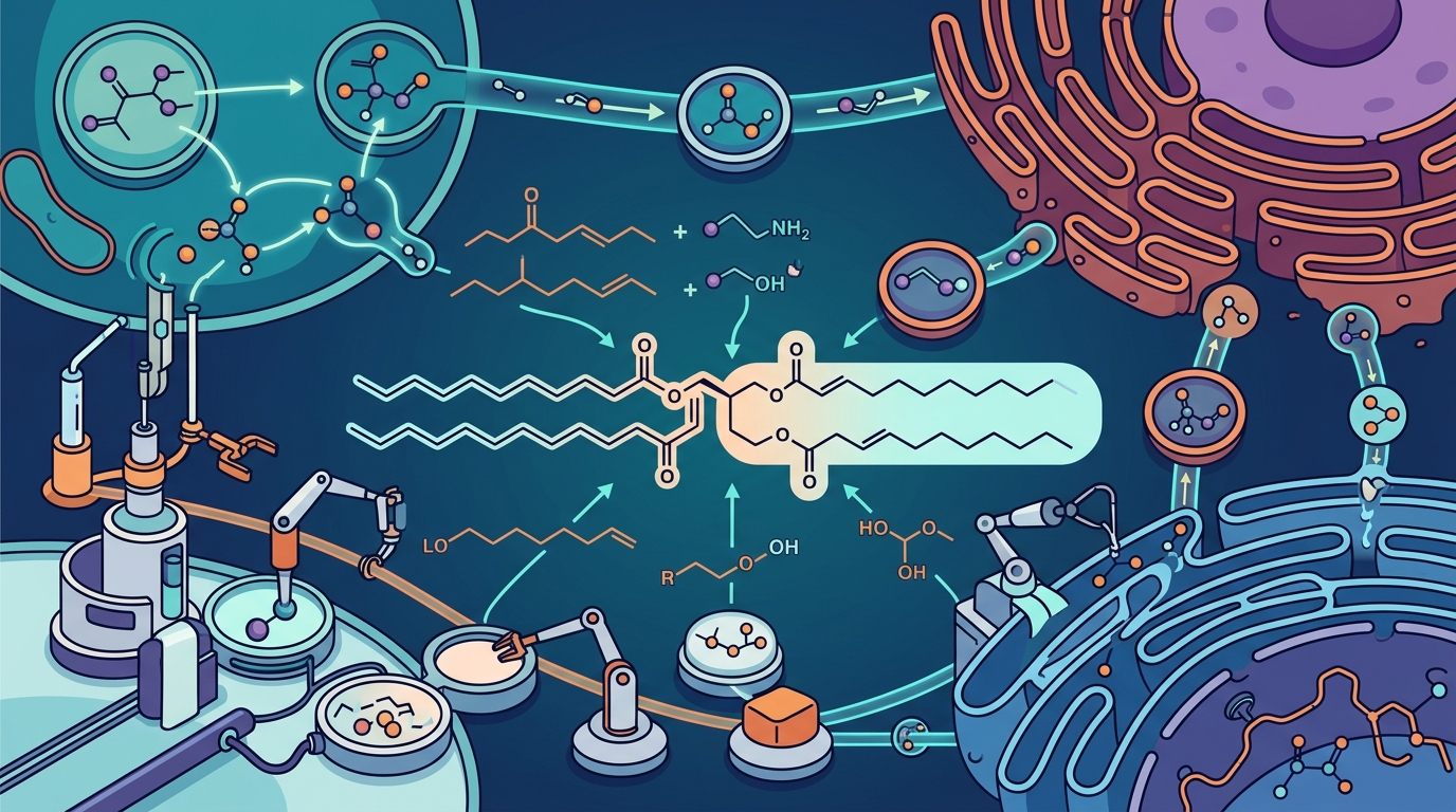
Step 1 — Acylation of DHAP (Peroxisome)
The pathway begins when the enzyme glyceronephosphate O-acyltransferase (GNPAT), also called DHAP-AT, attaches a long-chain fatty acyl-CoA to dihydroxyacetone phosphate (DHAP) at the sn-1 position, forming 1-acyl-DHAP. GNPAT is a peroxisomal matrix enzyme that faces the luminal side of the membrane and is considered the entry point for ether lipid biosynthesis.
Step 2 — Ether Bond Formation (Peroxisome)
Alkylglycerone phosphate synthase (AGPS), also called ADHAP-S, then replaces the acyl chain on 1-acyl-DHAP with a long-chain fatty alcohol, creating 1-O-alkyl-DHAP—the molecule that carries the characteristic ether bond. GNPAT and AGPS physically associate on the peroxisomal membrane, forming a functional complex that channels the intermediate directly from one active site to the next.
Step 3 — Reduction to Alkyl-Glycerophosphate (Peroxisome / ER)
Acyl/alkyl DHAP reductase (ADHAPR, also called PexRAP) reduces the ketone group of alkyl-DHAP to a hydroxyl, generating 1-O-alkyl-glycero-3-phosphate (AGP). This enzyme operates in both peroxisomes and the ER, providing flexibility in where the reaction takes place.
Steps 4–7 — Completion in the ER
The remaining steps mirror conventional phospholipid synthesis: a fatty acyl-CoA (often DHA or AA) is added at sn-2, a CDP-ethanolamine or CDP-choline head group is attached at sn-3, and finally a plasmalogen-specific desaturase (TMEM189/PEDS1) introduces the cis-double bond adjacent to the ether linkage, converting the alkyl intermediate into the mature vinyl-ether plasmalogen.
The Rate-Limiting Bottleneck: FAR1 and Fatty Alcohol Supply
Every plasmalogen molecule requires a fatty alcohol donor at Step 2. The enzyme fatty acyl-CoA reductase 1 (FAR1) generates these fatty alcohols by reducing long-chain acyl-CoAs in an NADPH-dependent reaction on the cytoplasmic face of the peroxisome. FAR1 is widely regarded as the rate-limiting enzyme for the entire pathway.
FAR1 activity directly controls how many fatty alcohol molecules are available for AGPS. When cellular plasmalogen levels are adequate, FAR1 protein is rapidly degraded; when levels fall, FAR1 stabilizes and production accelerates. This elegant feedback loop keeps plasmalogen homeostasis in check—but it also means that anything impairing FAR1 function or peroxisomal health will throttle the entire biosynthetic pipeline.
Feedback Regulation—How Cells Know When to Stop
Plasmalogen biosynthesis is spatiotemporally regulated by sensing plasmalogen concentration in the inner leaflet of the plasma membrane. When plasmalogen content in the inner leaflet is sufficient, FAR1 is tagged for proteasomal degradation, slowing fatty alcohol supply and, with it, overall plasmalogen output. Conversely, when membrane plasmalogens drop—due to oxidative degradation, increased demand, or impaired synthesis—FAR1 is stabilized, and production ramps up.
Additionally, TMEM189, the desaturase that inserts the final vinyl-ether bond, has been shown to influence FAR1 stability, adding a second layer of regulatory control at the terminal step.
Nutrients and Cofactors That Support Plasmalogen Production
Because plasmalogen biosynthesis depends on specific substrates, cofactors, and healthy organelle function, several dietary nutrients play supportive roles:
1. Omega-3 Fatty Acids (DHA and EPA)
DHA is the predominant PUFA found at the sn-2 position of brain ethanolamine plasmalogens. Adequate DHA intake—from fatty fish, fish oil, or algae oil—ensures the ER has an abundant supply of the acyl chains it needs to complete plasmalogen assembly. Research in animal models has demonstrated that EPA-enriched ethanolamine plasmalogen supplementation can significantly increase DHA content in the brain.
2. Choline and Ethanolamine
The polar head groups of plasmalogens are derived from ethanolamine or choline, both supplied through diet and endogenous synthesis. Eggs, liver, soybeans, and cruciferous vegetables are rich dietary sources. Choline also feeds into the CDP-choline pathway used for choline-plasmalogen assembly.
3. B Vitamins (B12, Folate, B6)
B vitamins support one-carbon metabolism and methylation reactions that maintain peroxisomal and mitochondrial function. Vitamin B12 and folate are especially important: impaired methylation can compromise membrane phospholipid remodeling and has been associated with lower plasmalogen levels in aging populations.
4. Iron and Copper
Several enzymes in the pathway—and in the broader peroxisomal beta-oxidation network—require metal cofactors. Iron, in particular, is needed for the desaturase step. Adequate but not excessive iron intake supports the catalytic machinery without amplifying oxidative stress.
5. NADPH (Supported by Niacin / Vitamin B3)
FAR1 is an NADPH-dependent reductase. NADPH regeneration depends on the pentose phosphate pathway and malic enzyme, both of which are influenced by niacin status and overall metabolic health.
6. Alkylglycerols (Dietary Precursors)
Alkylglycerols—found in shark liver oil, human breast milk, and bone marrow—can bypass the peroxisomal steps entirely and be converted to plasmalogens directly in the ER. This dietary shortcut is particularly interesting for individuals with compromised peroxisomal function.
7. Antioxidants (Vitamin E, Selenium, Polyphenols)
Because plasmalogens are themselves sacrificial antioxidants—their vinyl-ether bond scavenges reactive oxygen species—chronic oxidative stress accelerates their degradation. Dietary antioxidants help preserve existing plasmalogens, reducing the biosynthetic burden on peroxisomes.
Why Plasmalogen Production Declines with Age
Multiple lines of evidence now show that plasmalogen levels drop progressively after midlife. The decline is multifactorial:
- Reduced peroxisome biogenesis and protein import: Aging impairs the PEX-mediated protein import machinery, meaning fewer functional GNPAT and AGPS molecules reach the peroxisomal matrix.
- Increased oxidative degradation: Chronic oxidative stress in the aging brain breaks down existing plasmalogens faster than they can be replaced. Mitochondrial dysfunction further amplifies reactive oxygen species (ROS) production.
- Lower DHA synthesis and incorporation: The synthesis of DHA is itself reduced in aging, and its incorporation into ethanolamine plasmalogens declines in parallel.
- Up-regulation of degradative enzymes: Research indicates that the enzymes that break down ethanolamine plasmalogens increase with age, compounding the synthetic shortfall.
- Impaired pexophagy: Damaged peroxisomes accumulate when the cell's selective autophagy pathway (pexophagy) becomes less efficient, further reducing the pool of healthy, productive organelles.
This age-related decline in plasmalogen biosynthesis has been linked to neurodegenerative conditions including Alzheimer's disease, Parkinson's disease, and ALS, where postmortem analyses consistently reveal reduced plasmalogen levels and markers of peroxisomal dysfunction.
Key Takeaways
- Plasmalogen biosynthesis is a seven-step process that begins in peroxisomes and finishes in the endoplasmic reticulum.
- Three peroxisomal enzymes—GNPAT, AGPS, and FAR1—control the critical first stages, with FAR1 acting as the rate-limiting bottleneck.
- Cells regulate plasmalogen output through a feedback loop that senses plasmalogen levels in the inner leaflet of the plasma membrane and adjusts FAR1 stability accordingly.
- DHA, choline, ethanolamine, B vitamins, iron, and NADPH are among the key nutrients that feed into the biosynthetic machinery.
- Dietary alkylglycerols can bypass the peroxisomal steps, offering a potential workaround when peroxisomal function is compromised.
- Plasmalogen production declines with age due to reduced peroxisome biogenesis, increased oxidative stress, and impaired pexophagy—making nutritional and lifestyle support increasingly important over time.
Frequently Asked Questions
Where in the cell are plasmalogens made?
Plasmalogen biosynthesis starts inside peroxisomes, where the first two steps create the ether bond, and is completed in the endoplasmic reticulum (ER), where the sn-2 fatty acid and head group are added.
What is the rate-limiting enzyme in plasmalogen synthesis?
Fatty acyl-CoA reductase 1 (FAR1) is considered the rate-limiting enzyme. It produces the fatty alcohol substrates needed by AGPS to form the ether bond, and its protein stability is regulated by cellular plasmalogen levels.
Can you get plasmalogens from food?
Small amounts of preformed plasmalogens are found in shellfish, organ meats, and eggs. Alkylglycerols in shark liver oil and human breast milk can also be converted to plasmalogens in the ER, bypassing the peroxisomal steps. However, most of the body's plasmalogen supply comes from de novo synthesis in peroxisomes.
Why do plasmalogen levels drop with age?
Aging brings reduced peroxisome biogenesis, impaired protein import, increased oxidative degradation, lower DHA synthesis, up-regulated breakdown enzymes, and dysfunctional pexophagy—all of which reduce the net plasmalogen pool.
What nutrients help my body produce plasmalogens?
Key nutrients include omega-3 fatty acids (DHA, EPA), choline, ethanolamine, B vitamins (especially B12 and folate), iron, niacin (for NADPH), and dietary antioxidants like vitamin E. Alkylglycerols from marine sources can also serve as direct precursors.
What happens if peroxisomes are dysfunctional?
Peroxisomal disorders—whether inherited (such as Zellweger syndrome or RCDP) or acquired through aging—lead to severely impaired plasmalogen synthesis. This can manifest as neurological dysfunction, skeletal abnormalities, impaired myelination, and increased oxidative damage.
Sources and Further Reading
- Honsho M, Fujiki Y. Regulation of plasmalogen biosynthesis in mammalian cells and tissues. Brain Research Bulletin, 2023.
- Gu J, et al. Advances in the Biosynthetic Pathways and Application Potential of Plasmalogens in Medicine. Frontiers in Cell and Developmental Biology, 2020.
- Honsho M, et al. Plasmalogen biosynthesis is spatiotemporally regulated by sensing plasmalogens in the inner leaflet of plasma membranes. Scientific Reports, 2017.
- Dorninger F, et al. Plasmalogens and Chronic Inflammatory Diseases. Frontiers in Physiology, 2021.
- Huang J, et al. Peroxisomes in Aging: Guardians of Cellular Resilience and Function. Cells, 2025.