
Plasmalogens are among the most abundant yet least understood lipids in the human body. Found in every cell membrane—and concentrated in the brain, heart, and immune system—these specialized phospholipids influence everything from neuronal signaling to oxidative defense. This ultimate guide explores their chemistry, biosynthesis, tissue distribution, and the growing body of research linking plasmalogen levels to aging, neurodegeneration, and chronic disease.
Defining Plasmalogens: Chemistry and Classification
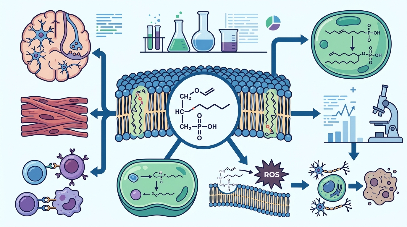
Plasmalogens are a class of glycerophospholipids distinguished by a vinyl-ether bond at the sn-1 position of the glycerol backbone. Unlike typical phospholipids that carry ester bonds at both sn-1 and sn-2 positions, plasmalogens feature this unique alkenyl linkage, which gives them distinct biophysical and biochemical properties.
Structurally, plasmalogens carry a fatty alcohol linked through the vinyl-ether bond at sn-1, while the sn-2 position is typically enriched in polyunsaturated fatty acids (PUFAs) such as docosahexaenoic acid (DHA), arachidonic acid (AA), and eicosapentaenoic acid (EPA). The head group at sn-3 is most commonly ethanolamine (forming plasmenylethanolamine, or PlsEtn) or choline (forming plasmenylcholine, or PlsCho).
Within the broader category of ether lipids, two subtypes exist: plasmanyl lipids (with an alkyl ether bond) and plasmenyl lipids, also known as plasmalogens (with a vinyl-ether bond). It is the vinyl-ether linkage that sets plasmalogens apart and confers their antioxidant capacity and membrane-modulating behavior.
How the Body Makes Plasmalogens: Peroxisomes and Beyond
Plasmalogen biosynthesis is a multi-organelle process that begins in the peroxisome and is completed in the endoplasmic reticulum. The first two enzymatic steps—catalyzed by glyceronephosphate O-acyltransferase (GNPAT) and alkyldihydroxyacetone phosphate synthase (AGPS)—occur exclusively within the peroxisome. This is why peroxisomal health is a prerequisite for adequate plasmalogen levels.
The fatty alcohol substrate at sn-1 is produced by fatty acyl-CoA reductase 1 (FAR1), which resides on the peroxisomal membrane. The intermediate is then shuttled to the endoplasmic reticulum, where the final vinyl-ether bond is introduced by the enzyme plasmanylethanolamine desaturase (PEDS1, encoded by TMEM189). This last step converts the plasmanyl precursor into a mature plasmenyl (plasmalogen) species.
Because this biosynthetic route is nonredundant—no alternative pathway exists in mammals—any disruption in peroxisomal function directly reduces plasmalogen availability throughout the body. Genetic defects at these steps cause Rhizomelic Chondrodysplasia Punctata (RCDP), a severe malformation syndrome affecting bone, brain, lens, lung, kidney, and heart.
Tissue Distribution: Where Plasmalogens Concentrate
Plasmalogens are not uniformly distributed. They are most abundant in tissues with high metabolic or signaling demands. In total, plasmalogens account for approximately 15–20 mol% of all phospholipids in mammalian tissues, but certain cell types contain far higher proportions.
- Brain and nervous system: Approximately 20% of brain glycerophospholipids are plasmalogens. In the myelin sheath specifically, ethanolamine plasmalogens make up roughly 70% of all ethanolamine glycerophospholipids.
- Heart: Choline plasmalogens represent 30–40% of choline glycerophospholipids in cardiac tissue, making the heart one of the most plasmalogen-rich organs.
- Immune cells: Neutrophils, macrophages, and other immune cells carry substantial plasmalogen loads, reflecting these lipids' roles in membrane signaling and inflammatory modulation.
- Red blood cells and lipoproteins: Plasmalogens are also found in erythrocyte membranes and circulating HDL and LDL particles.
Role in Cell Membrane Structure and Dynamics
Plasmalogens influence the physical architecture of cell membranes in several measurable ways. The absence of the carbonyl oxygen at sn-1 reduces the polarity at the membrane interface, promoting tighter intermolecular hydrogen bonding between adjacent phospholipids. This alters membrane packing density, fluidity, thickness, and lateral pressure profiles.
These biophysical effects are not trivial. Plasmalogens are critical for the organization and stability of lipid raft microdomains—cholesterol-rich membrane regions that serve as platforms for receptor clustering, signal transduction, and vesicular trafficking. They also facilitate the formation of non-lamellar (hexagonal II) membrane phases, which are essential for membrane fusion events such as synaptic vesicle release.
By modulating membrane curvature and dynamics, plasmalogens support the proper function of integral membrane proteins, ion channels, and ionotropic receptors—making them indispensable for cellular communication throughout the nervous and cardiovascular systems.
Plasmalogens as Endogenous Antioxidants
One of the most studied functions of plasmalogens is their role as sacrificial antioxidants. The vinyl-ether bond at sn-1 is highly susceptible to oxidation by reactive oxygen species (ROS), including singlet oxygen and peroxyl radicals. When ROS attack the vinyl-ether bond, the plasmalogen molecule is preferentially oxidized instead of more critical membrane components such as polyunsaturated fatty acids and membrane proteins.
This sacrificial mechanism is especially significant in tissues with high metabolic rates or elevated oxidative stress exposure, such as the brain, retina, and heart. Beyond direct radical scavenging, plasmalogens also modulate downstream inflammatory responses and cell survival pathways, linking their antioxidant function to broader anti-inflammatory effects at the cellular level.
The practical implication: as plasmalogen levels decline—whether through aging, peroxisomal dysfunction, or disease—the cell membrane loses a primary line of antioxidant defense, leaving PUFAs and membrane proteins increasingly vulnerable to oxidative degradation.
Brain Health, Myelin, and Cognitive Function
The nervous system depends on plasmalogens more than almost any other tissue. These lipids are essential structural components of myelin, the insulating sheath around nerve axons that enables rapid signal conduction. In myelin, ethanolamine plasmalogens represent up to 70% of ethanolamine glycerophospholipids—an extraordinary concentration that underscores their structural necessity.
Beyond myelin integrity, plasmalogens support synaptic function. They are enriched in synaptic vesicle membranes, where their ability to promote membrane fusion facilitates neurotransmitter release. DHA-containing plasmalogens are the most abundant plasmalogen species in cerebral cortex membranes, linking their biology directly to the well-documented neuroprotective effects of omega-3 fatty acids.
Research also suggests that plasmalogens may modulate ion channels and receptor activity, influencing neurotransmission efficiency. A mouse study demonstrated that plasmalogens can eliminate aging-associated synaptic defects, pointing to a direct role in maintaining cognitive performance as the brain ages.
Disease Associations: From Alzheimer's to Cardiovascular Disorders
Reduced plasmalogen levels have been documented across a surprisingly broad spectrum of human diseases. The most extensively studied association is with Alzheimer's disease (AD). Research has shown dramatic decreases in brain plasmalogen content—up to 40 mol% reduction in white matter—at very early stages of AD. In gray matter, the deficiency correlates with disease severity, progressing from roughly 10% reduction in very mild dementia to about 30% in severe cases.
Longitudinal epidemiological data add further weight: a decrease in the plasmalogen index over time was associated with higher odds of converting from normal cognition to mild cognitive impairment or AD, while a higher baseline plasmalogen index was protective. Circulating ethanolamine plasmalogen levels are significantly decreased in serum from AD subjects at all stages of dementia.
The disease associations extend well beyond neurodegeneration:
- Parkinson's disease and multiple sclerosis: Reduced brain plasmalogen levels are consistently reported.
- Cardiovascular disease: Altered plasmalogen metabolism is described in heart disease.
- Metabolic disorders: Diabetes and obesity show plasmalogen abnormalities.
- Psychiatric conditions: Depression, bipolar disorder, and schizophrenia have all been linked to lower plasmalogen levels.
- Respiratory disease: Reduced plasmalogens are observed in COPD patients who smoke.
- Cancer: Altered plasmalogen metabolism has been reported in breast, ovarian, lung, and gastrointestinal cancers.
- Infectious disease: COVID-19 patients show significant reductions in plasmalogen levels, with strong correlations to symptom severity.
Whether plasmalogen deficiency is a cause, consequence, or amplifier of these conditions remains an active area of investigation. Current evidence supports the view that plasmalogen deficiency increases susceptibility to neurodegeneration and other pathologies, potentially interacting with other risk factors to initiate or worsen disease.
Aging, Longevity, and Plasmalogen Decline
Circulating plasmalogen levels decrease with age. Because plasmalogen biosynthesis depends on peroxisomal function—which itself declines during aging—older adults progressively lose the capacity to maintain adequate plasmalogen pools. This age-related decline is observed in both serum measurements and postmortem brain tissue analyses.
Intriguingly, studies of human centenarians and model organisms such as Caenorhabditis elegans have shed light on the relationship between plasmalogens and longevity, although a direct causal link requires further investigation. The convergence of plasmalogen loss with increased oxidative stress, mitochondrial dysfunction, and chronic low-grade inflammation during aging suggests that plasmalogen depletion may be a unifying biochemical factor underlying multiple age-related pathologies.
Serum plasmalogens have also been shown to positively correlate with high-density lipoprotein (HDL) levels—a well-established marker of cardiovascular health—adding another dimension to the aging-plasmalogen connection.
Measuring Plasmalogens: Lipidomics and Biomarker Science
Advances in mass spectrometry-based lipidomics have made it possible to quantify individual plasmalogen species from blood plasma, serum, erythrocytes, platelets, and peripheral blood mononuclear cells. However, challenges remain. Published isolation protocols vary, and mass overlaps between lipid species can complicate accurate measurement.
The plasmalogen index—typically expressed as a ratio of plasmalogen to phosphatidylethanolamine species—has emerged as a promising composite biomarker. Reduced indices of plasmalogen biosynthesis and remodeling have been significantly correlated with elevated cerebrospinal fluid concentrations of total tau, a biomarker of neurodegeneration.
As lipidomic technologies mature, plasmalogens may become valuable diagnostic and prognostic biomarkers for vascular disease, metabolic syndrome, neurodegeneration, and even cancer. Standardization of analytical methods across laboratories remains an important prerequisite for clinical adoption.
Plasmalogen Replacement Therapy: Current Research
Plasmalogen Replacement Therapy (PRT) is an emerging therapeutic approach that aims to restore depleted plasmalogen levels in the body. Several strategies are under investigation:
- Oral precursors: Alkyl-diacylglycerol compounds, particularly DHA-containing alkyl-diacylglycerol (DHA-AAG), have been shown to increase circulating DHA-plasmalogen levels significantly. In one clinical study, a single dose at 100 mg/kg increased circulating plasmalogen levels by 80% within 24 hours in healthy subjects.
- Scallop-derived plasmalogens: Research has shown that 1 mg/day of scallop-derived plasmalogens improved memory function in patients with mild AD, though larger trials are needed.
- Nanomedicine approaches: Lipid nanoparticles and advanced drug delivery systems are being explored to overcome the challenge of delivering plasmalogens across the blood-brain barrier, which remains a substantial obstacle for oral formulations targeting CNS disorders.
Despite promising early results, recent clinical studies have not always demonstrated significant cognitive improvements after oral plasmalogen administration, potentially due to insufficient dosage or limited bioavailability. Future research is focused on optimizing delivery methods, including intranasal routes and targeted nanoparticle systems.
Key Takeaways
- Plasmalogens are vinyl-ether-bonded glycerophospholipids that make up approximately 15–20% of total phospholipids in human tissues.
- Their biosynthesis begins exclusively in peroxisomes—peroxisomal health is essential for maintaining plasmalogen levels.
- Plasmalogens are heavily concentrated in the brain (especially myelin), heart, and immune cells.
- They serve as sacrificial antioxidants, protecting membranes from reactive oxygen species.
- They are critical for membrane fluidity, lipid raft organization, and synaptic vesicle fusion.
- Plasmalogen deficiency is associated with Alzheimer's disease, Parkinson's disease, cardiovascular disease, metabolic disorders, psychiatric conditions, and more.
- Levels decline naturally with aging, paralleling increased disease risk.
- Mass spectrometry-based lipidomics enables measurement, but standardization is still needed.
- Plasmalogen Replacement Therapy is an active area of research with several promising preclinical and early clinical results.
Frequently Asked Questions
What exactly are plasmalogens?
Plasmalogens are a subclass of glycerophospholipids defined by a vinyl-ether bond at the sn-1 position of their glycerol backbone. They are abundant in cell membranes—particularly in the brain, heart, and immune system—and play roles in membrane structure, antioxidant defense, and cell signaling.
How are plasmalogens different from regular phospholipids?
Standard phospholipids have ester bonds at both sn-1 and sn-2 positions. Plasmalogens replace the sn-1 ester bond with a vinyl-ether linkage. This structural difference gives plasmalogens unique properties: stronger intermolecular hydrogen bonding, the ability to act as antioxidants, and distinct effects on membrane curvature and dynamics.
Where are plasmalogens made in the body?
The initial and rate-limiting steps of plasmalogen synthesis occur in peroxisomes. The pathway then continues in the endoplasmic reticulum, where the final vinyl-ether bond is formed by the enzyme PEDS1 (TMEM189). There is no alternative biosynthetic route in mammals.
Why do plasmalogen levels decline with age?
Peroxisomal function decreases during aging, reducing the body's capacity to synthesize new plasmalogens. At the same time, increased oxidative stress with aging accelerates the consumption of existing plasmalogens, compounding the decline.
Are plasmalogens linked to Alzheimer's disease?
Yes. Multiple studies have documented reduced plasmalogen levels in the brains and blood of Alzheimer's disease patients, with the magnitude of deficiency correlating with disease severity. Whether this deficiency is a cause, consequence, or amplifying factor of AD remains under active investigation, but growing evidence supports a causal contribution.
Can you get plasmalogens from food?
The body must synthesize most of its own plasmalogens through the peroxisomal pathway. While some dietary sources such as scallops and certain shellfish contain plasmalogens, these are largely broken down during digestion. Current research focuses on oral precursor compounds that can be absorbed and converted to plasmalogens within the body.
What is Plasmalogen Replacement Therapy?
Plasmalogen Replacement Therapy (PRT) uses precursor molecules or plasmalogen-containing compounds to raise depleted levels in the body. Several approaches, including DHA-containing alkyl-diacylglycerol and nanomedicine-based delivery systems, are under investigation for neurodegenerative, cardiovascular, and metabolic diseases.
How are plasmalogen levels measured?
Plasmalogen levels are most accurately measured using mass spectrometry-based lipidomics from blood samples (plasma, serum, or erythrocytes). A composite metric called the plasmalogen index can be calculated as a ratio of plasmalogen to phosphatidylethanolamine species and has shown correlations with disease biomarkers such as CSF tau.